Manganese »
PDB 2ypq-3au8 »
3a6d »
Manganese in PDB 3a6d: Creatininase Complexed with 1-Methylguanidine
Enzymatic activity of Creatininase Complexed with 1-Methylguanidine
All present enzymatic activity of Creatininase Complexed with 1-Methylguanidine:
3.5.2.10;
3.5.2.10;
Protein crystallography data
The structure of Creatininase Complexed with 1-Methylguanidine, PDB code: 3a6d
was solved by
Y.Nakajima,
K.Yamashita,
K.Ito,
T.Yoshimoto,
with X-Ray Crystallography technique. A brief refinement statistics is given in the table below:
| Resolution Low / High (Å) | 20.00 / 1.90 |
| Space group | P 21 21 21 |
| Cell size a, b, c (Å), α, β, γ (°) | 102.400, 152.700, 167.500, 90.00, 90.00, 90.00 |
| R / Rfree (%) | 19.2 / 20.7 |
Other elements in 3a6d:
The structure of Creatininase Complexed with 1-Methylguanidine also contains other interesting chemical elements:
| Zinc | (Zn) | 6 atoms |
Manganese Binding Sites:
The binding sites of Manganese atom in the Creatininase Complexed with 1-Methylguanidine
(pdb code 3a6d). This binding sites where shown within
5.0 Angstroms radius around Manganese atom.
In total 6 binding sites of Manganese where determined in the Creatininase Complexed with 1-Methylguanidine, PDB code: 3a6d:
Jump to Manganese binding site number: 1; 2; 3; 4; 5; 6;
In total 6 binding sites of Manganese where determined in the Creatininase Complexed with 1-Methylguanidine, PDB code: 3a6d:
Jump to Manganese binding site number: 1; 2; 3; 4; 5; 6;
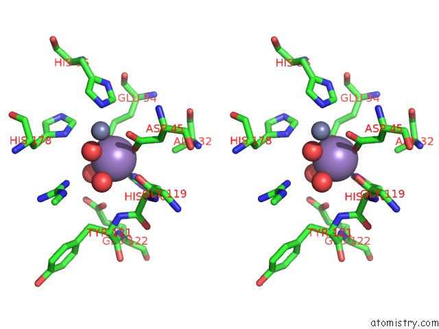
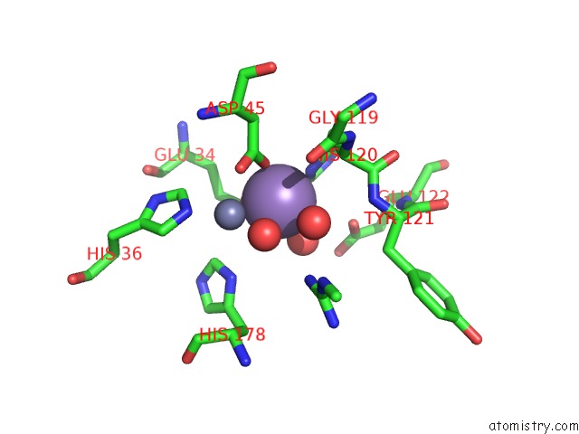
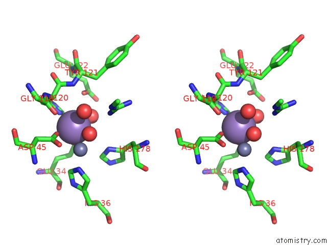
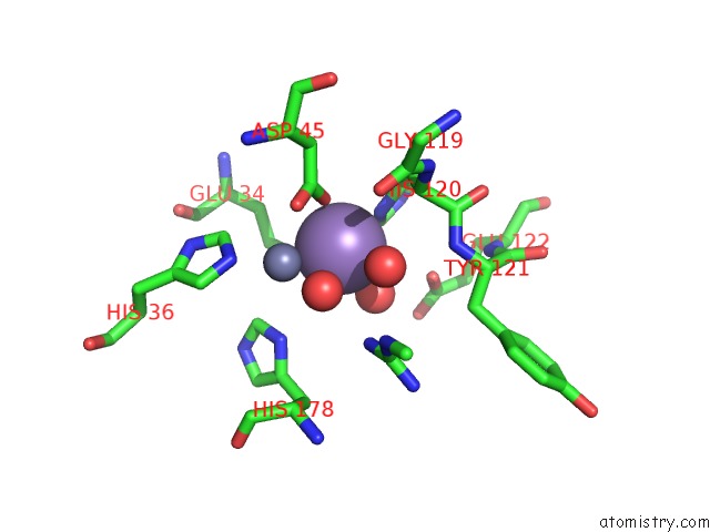
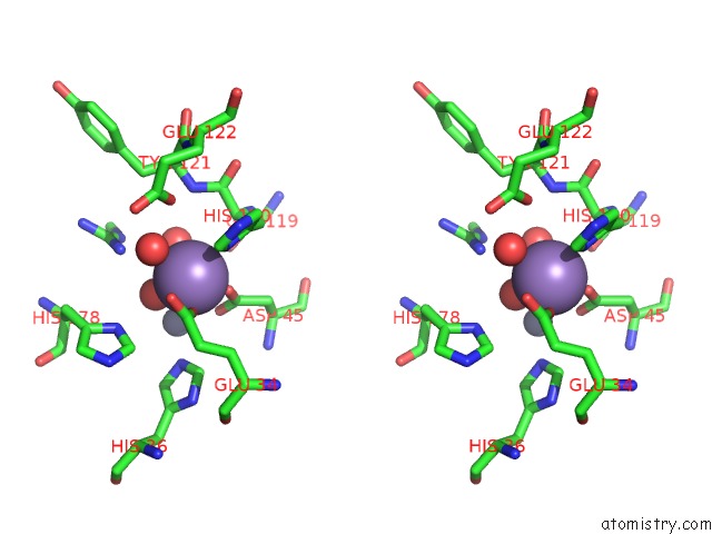
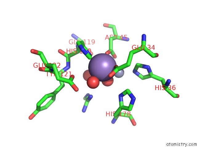
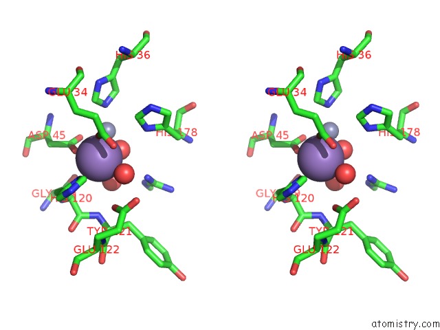
Manganese binding site 1 out of 6 in 3a6d
Go back to
Manganese binding site 1 out
of 6 in the Creatininase Complexed with 1-Methylguanidine
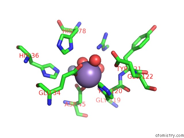
Mono view

Stereo pair view
Mono view
Stereo pair view
A full contact list of Manganese with other atoms in the Mn binding
site number 1 of Creatininase Complexed with 1-Methylguanidine within 5.0Å range:
|
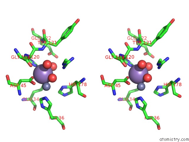
Manganese binding site 2 out of 6 in 3a6d
Go back to
Manganese binding site 2 out
of 6 in the Creatininase Complexed with 1-Methylguanidine
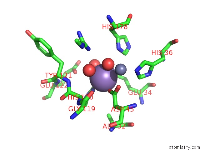
Mono view
Stereo pair view
Mono view
Stereo pair view
A full contact list of Manganese with other atoms in the Mn binding
site number 2 of Creatininase Complexed with 1-Methylguanidine within 5.0Å range:
|
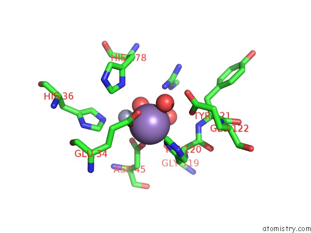
Manganese binding site 3 out of 6 in 3a6d
Go back to
Manganese binding site 3 out
of 6 in the Creatininase Complexed with 1-Methylguanidine
Mono view
Stereo pair view
Mono view
Stereo pair view
A full contact list of Manganese with other atoms in the Mn binding
site number 3 of Creatininase Complexed with 1-Methylguanidine within 5.0Å range:
|
Manganese binding site 4 out of 6 in 3a6d
Go back to
Manganese binding site 4 out
of 6 in the Creatininase Complexed with 1-Methylguanidine
Mono view
Stereo pair view
Mono view
Stereo pair view
A full contact list of Manganese with other atoms in the Mn binding
site number 4 of Creatininase Complexed with 1-Methylguanidine within 5.0Å range:
|
Manganese binding site 5 out of 6 in 3a6d
Go back to
Manganese binding site 5 out
of 6 in the Creatininase Complexed with 1-Methylguanidine
Mono view
Stereo pair view
Mono view
Stereo pair view
A full contact list of Manganese with other atoms in the Mn binding
site number 5 of Creatininase Complexed with 1-Methylguanidine within 5.0Å range:
|
Manganese binding site 6 out of 6 in 3a6d
Go back to
Manganese binding site 6 out
of 6 in the Creatininase Complexed with 1-Methylguanidine
Mono view
Stereo pair view
Mono view
Stereo pair view
A full contact list of Manganese with other atoms in the Mn binding
site number 6 of Creatininase Complexed with 1-Methylguanidine within 5.0Å range:
|
Reference:
K.Yamashita,
Y.Nakajima,
H.Matsushita,
Y.Nishiya,
R.Yamazawa,
Y.F.Wu,
F.Matsubara,
H.Oyama,
K.Ito,
T.Yoshimoto.
Substitution of GLU122 By Glutamine Revealed the Function of the Second Water Molecule As A Proton Donor in the Binuclear Metal Enzyme Creatininase J.Mol.Biol. V. 396 1081 2010.
ISSN: ISSN 0022-2836
PubMed: 20043918
DOI: 10.1016/J.JMB.2009.12.045
Page generated: Sat Aug 16 11:18:48 2025
ISSN: ISSN 0022-2836
PubMed: 20043918
DOI: 10.1016/J.JMB.2009.12.045
Last articles
Mn in 4QNJMn in 4QKF
Mn in 4QKN
Mn in 4Q42
Mn in 4Q41
Mn in 4QAX
Mn in 4QH9
Mn in 4QAG
Mn in 4Q7I
Mn in 4Q40