Manganese »
PDB 4php-4qsf »
4q42 »
Manganese in PDB 4q42: Crystal Structure of Schistosoma Mansoni Arginase in Complex with L- Ornithine
Enzymatic activity of Crystal Structure of Schistosoma Mansoni Arginase in Complex with L- Ornithine
All present enzymatic activity of Crystal Structure of Schistosoma Mansoni Arginase in Complex with L- Ornithine:
3.5.3.1;
3.5.3.1;
Protein crystallography data
The structure of Crystal Structure of Schistosoma Mansoni Arginase in Complex with L- Ornithine, PDB code: 4q42
was solved by
Y.Hai,
J.E.Edwards,
M.C.Van Zandt,
K.F.Hoffmann,
D.W.Christianson,
with X-Ray Crystallography technique. A brief refinement statistics is given in the table below:
| Resolution Low / High (Å) | 49.49 / 2.05 |
| Space group | P 21 3 |
| Cell size a, b, c (Å), α, β, γ (°) | 178.447, 178.447, 178.447, 90.00, 90.00, 90.00 |
| R / Rfree (%) | 17.4 / 20.8 |
Manganese Binding Sites:
The binding sites of Manganese atom in the Crystal Structure of Schistosoma Mansoni Arginase in Complex with L- Ornithine
(pdb code 4q42). This binding sites where shown within
5.0 Angstroms radius around Manganese atom.
In total 8 binding sites of Manganese where determined in the Crystal Structure of Schistosoma Mansoni Arginase in Complex with L- Ornithine, PDB code: 4q42:
Jump to Manganese binding site number: 1; 2; 3; 4; 5; 6; 7; 8;
In total 8 binding sites of Manganese where determined in the Crystal Structure of Schistosoma Mansoni Arginase in Complex with L- Ornithine, PDB code: 4q42:
Jump to Manganese binding site number: 1; 2; 3; 4; 5; 6; 7; 8;
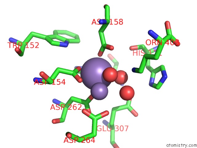
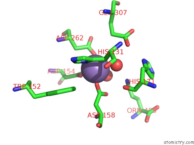
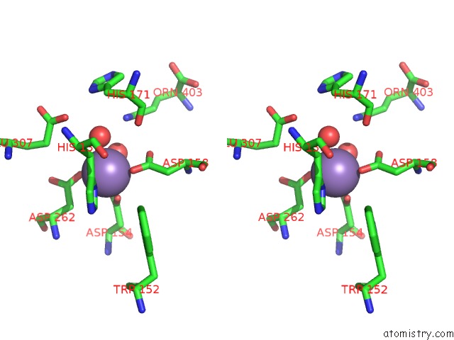
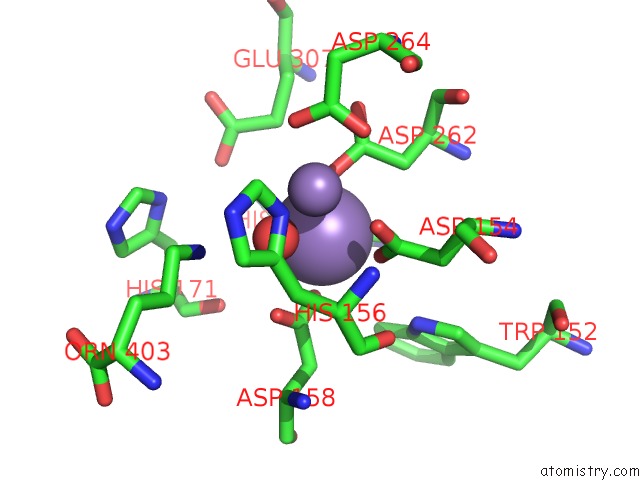
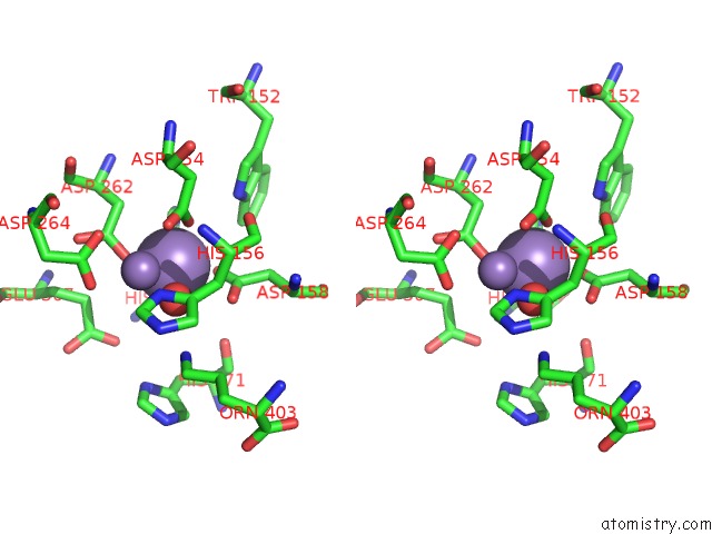
Manganese binding site 1 out of 8 in 4q42
Go back to
Manganese binding site 1 out
of 8 in the Crystal Structure of Schistosoma Mansoni Arginase in Complex with L- Ornithine
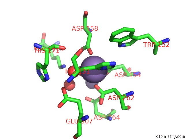
Mono view
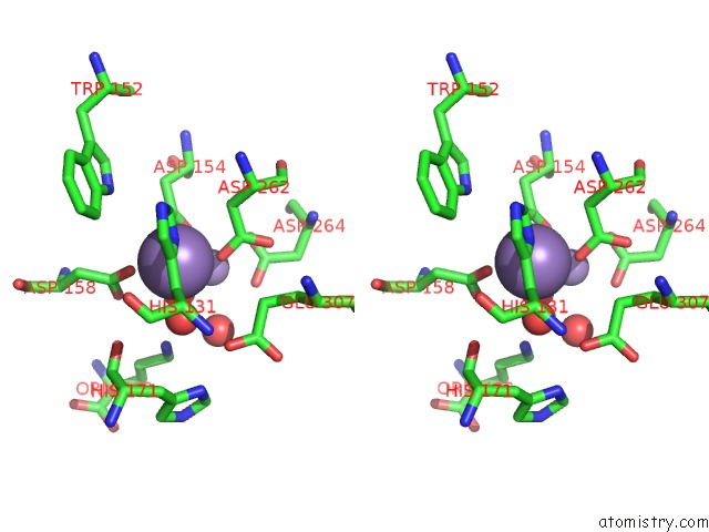
Stereo pair view
Mono view
Stereo pair view
A full contact list of Manganese with other atoms in the Mn binding
site number 1 of Crystal Structure of Schistosoma Mansoni Arginase in Complex with L- Ornithine within 5.0Å range:
|
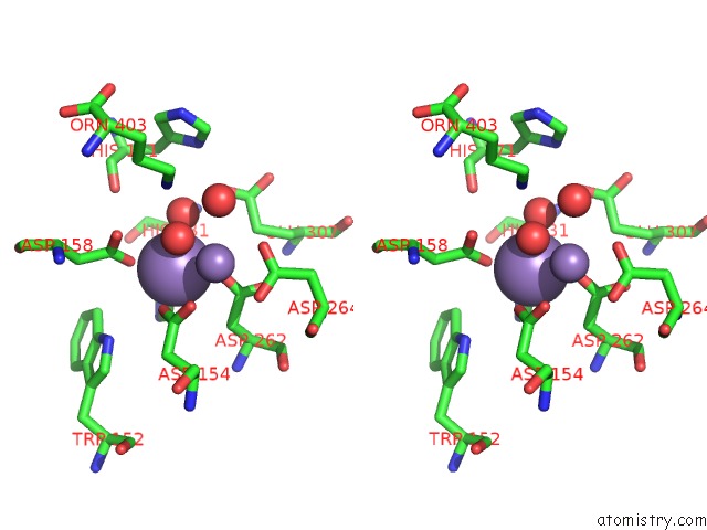
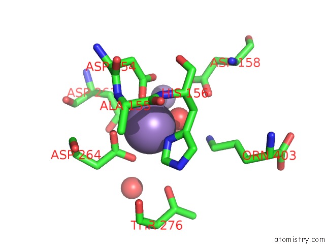
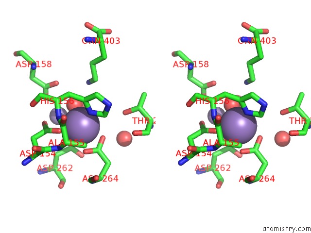
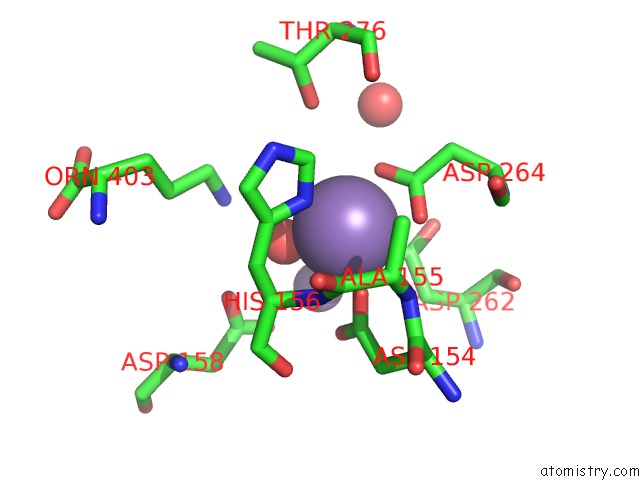
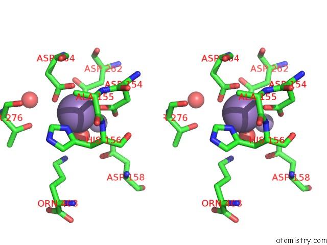
Manganese binding site 2 out of 8 in 4q42
Go back to
Manganese binding site 2 out
of 8 in the Crystal Structure of Schistosoma Mansoni Arginase in Complex with L- Ornithine
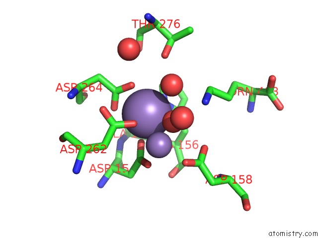
Mono view
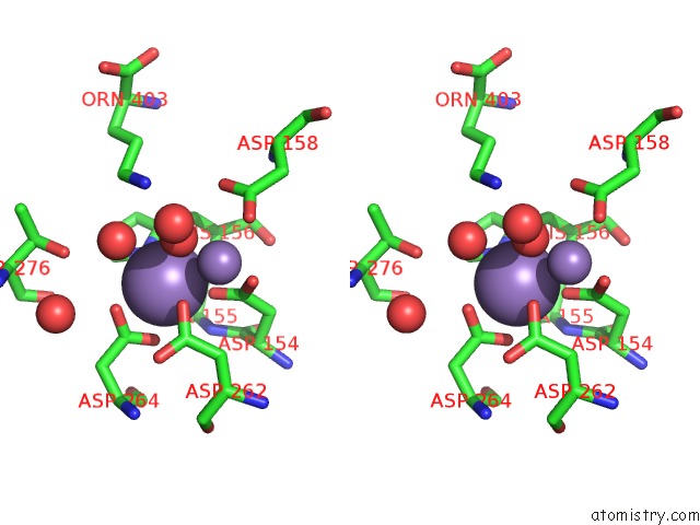
Stereo pair view
Mono view
Stereo pair view
A full contact list of Manganese with other atoms in the Mn binding
site number 2 of Crystal Structure of Schistosoma Mansoni Arginase in Complex with L- Ornithine within 5.0Å range:
|
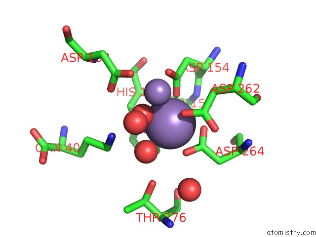
Manganese binding site 3 out of 8 in 4q42
Go back to
Manganese binding site 3 out
of 8 in the Crystal Structure of Schistosoma Mansoni Arginase in Complex with L- Ornithine
Mono view
Stereo pair view
Mono view
Stereo pair view
A full contact list of Manganese with other atoms in the Mn binding
site number 3 of Crystal Structure of Schistosoma Mansoni Arginase in Complex with L- Ornithine within 5.0Å range:
|
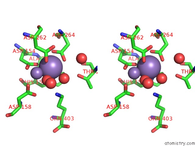
Manganese binding site 4 out of 8 in 4q42
Go back to
Manganese binding site 4 out
of 8 in the Crystal Structure of Schistosoma Mansoni Arginase in Complex with L- Ornithine
Mono view
Stereo pair view
Mono view
Stereo pair view
A full contact list of Manganese with other atoms in the Mn binding
site number 4 of Crystal Structure of Schistosoma Mansoni Arginase in Complex with L- Ornithine within 5.0Å range:
|
Manganese binding site 5 out of 8 in 4q42
Go back to
Manganese binding site 5 out
of 8 in the Crystal Structure of Schistosoma Mansoni Arginase in Complex with L- Ornithine
Mono view
Stereo pair view
Mono view
Stereo pair view
A full contact list of Manganese with other atoms in the Mn binding
site number 5 of Crystal Structure of Schistosoma Mansoni Arginase in Complex with L- Ornithine within 5.0Å range:
|
Manganese binding site 6 out of 8 in 4q42
Go back to
Manganese binding site 6 out
of 8 in the Crystal Structure of Schistosoma Mansoni Arginase in Complex with L- Ornithine
Mono view
Stereo pair view
Mono view
Stereo pair view
A full contact list of Manganese with other atoms in the Mn binding
site number 6 of Crystal Structure of Schistosoma Mansoni Arginase in Complex with L- Ornithine within 5.0Å range:
|
Manganese binding site 7 out of 8 in 4q42
Go back to
Manganese binding site 7 out
of 8 in the Crystal Structure of Schistosoma Mansoni Arginase in Complex with L- Ornithine
Mono view
Stereo pair view
Mono view
Stereo pair view
A full contact list of Manganese with other atoms in the Mn binding
site number 7 of Crystal Structure of Schistosoma Mansoni Arginase in Complex with L- Ornithine within 5.0Å range:
|
Manganese binding site 8 out of 8 in 4q42
Go back to
Manganese binding site 8 out
of 8 in the Crystal Structure of Schistosoma Mansoni Arginase in Complex with L- Ornithine
Mono view
Stereo pair view
Mono view
Stereo pair view
A full contact list of Manganese with other atoms in the Mn binding
site number 8 of Crystal Structure of Schistosoma Mansoni Arginase in Complex with L- Ornithine within 5.0Å range:
|
Reference:
Y.Hai,
J.E.Edwards,
M.C.Van Zandt,
K.F.Hoffmann,
D.W.Christianson.
Crystal Structure of Schistosoma Mansoni Arginase, A Potential Drug Target For the Treatment of Schistosomiasis. Biochemistry V. 53 4671 2014.
ISSN: ISSN 0006-2960
PubMed: 25007099
DOI: 10.1021/BI5004519
Page generated: Sat Oct 5 20:57:20 2024
ISSN: ISSN 0006-2960
PubMed: 25007099
DOI: 10.1021/BI5004519
Last articles
K in 3MMXK in 3MZ7
K in 3MZ4
K in 3MZ3
K in 3MZ6
K in 3MEN
K in 3MLB
K in 3MIO
K in 3MIJ
K in 3MD7