Manganese »
PDB 2a7a-2axt »
2amh »
Manganese in PDB 2amh: Crystal Structure of Maf-Like Protein TBRU21784AAA From T.Brucei
Protein crystallography data
The structure of Crystal Structure of Maf-Like Protein TBRU21784AAA From T.Brucei, PDB code: 2amh
was solved by
J.Caruthers,
E.Merritt,
Structural Genomics Of Pathogenic Protozoaconsortium (Sgpp),
with X-Ray Crystallography technique. A brief refinement statistics is given in the table below:
| Resolution Low / High (Å) | 18.00 / 2.00 |
| Space group | I 4 |
| Cell size a, b, c (Å), α, β, γ (°) | 97.068, 97.068, 48.894, 90.00, 90.00, 90.00 |
| R / Rfree (%) | 19.7 / 23.1 |
Manganese Binding Sites:
The binding sites of Manganese atom in the Crystal Structure of Maf-Like Protein TBRU21784AAA From T.Brucei
(pdb code 2amh). This binding sites where shown within
5.0 Angstroms radius around Manganese atom.
In total 3 binding sites of Manganese where determined in the Crystal Structure of Maf-Like Protein TBRU21784AAA From T.Brucei, PDB code: 2amh:
Jump to Manganese binding site number: 1; 2; 3;
In total 3 binding sites of Manganese where determined in the Crystal Structure of Maf-Like Protein TBRU21784AAA From T.Brucei, PDB code: 2amh:
Jump to Manganese binding site number: 1; 2; 3;
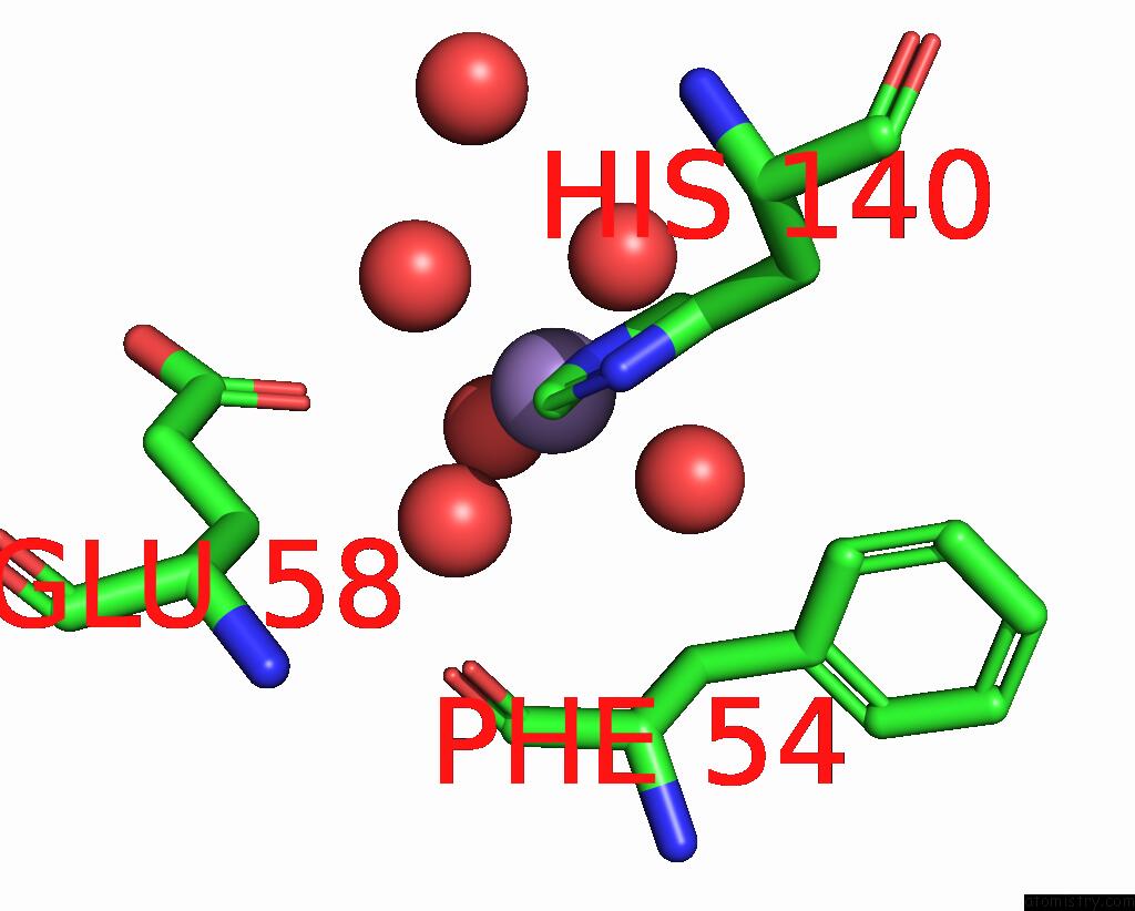
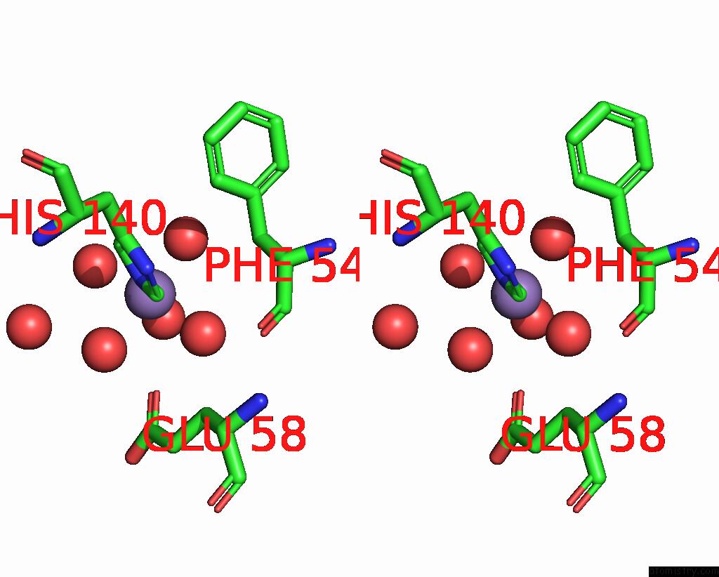
Manganese binding site 1 out of 3 in 2amh
Go back to
Manganese binding site 1 out
of 3 in the Crystal Structure of Maf-Like Protein TBRU21784AAA From T.Brucei
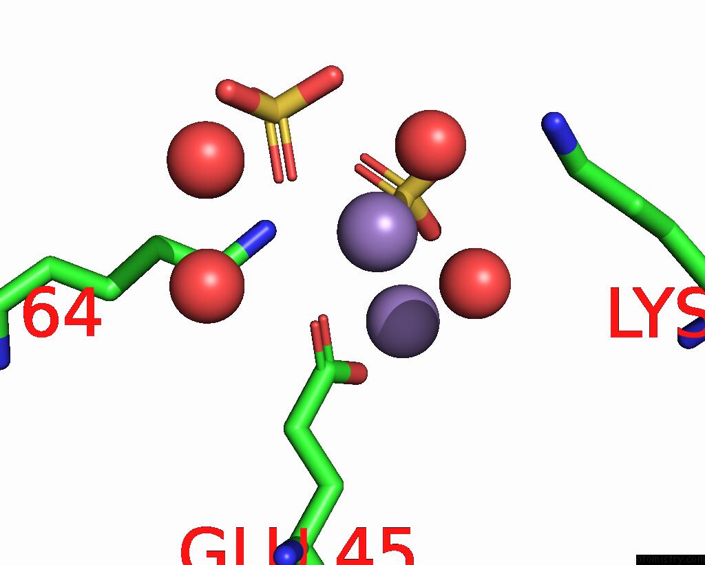
Mono view
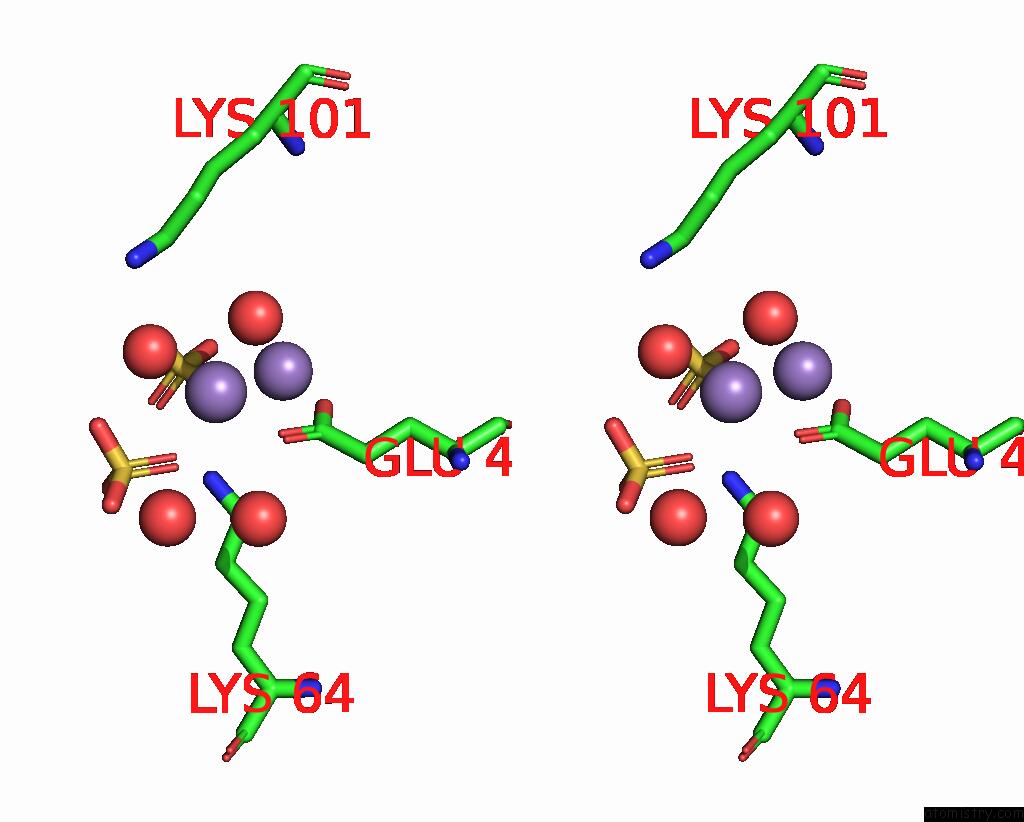
Stereo pair view
Mono view
Stereo pair view
A full contact list of Manganese with other atoms in the Mn binding
site number 1 of Crystal Structure of Maf-Like Protein TBRU21784AAA From T.Brucei within 5.0Å range:
|
Manganese binding site 2 out of 3 in 2amh
Go back to
Manganese binding site 2 out
of 3 in the Crystal Structure of Maf-Like Protein TBRU21784AAA From T.Brucei
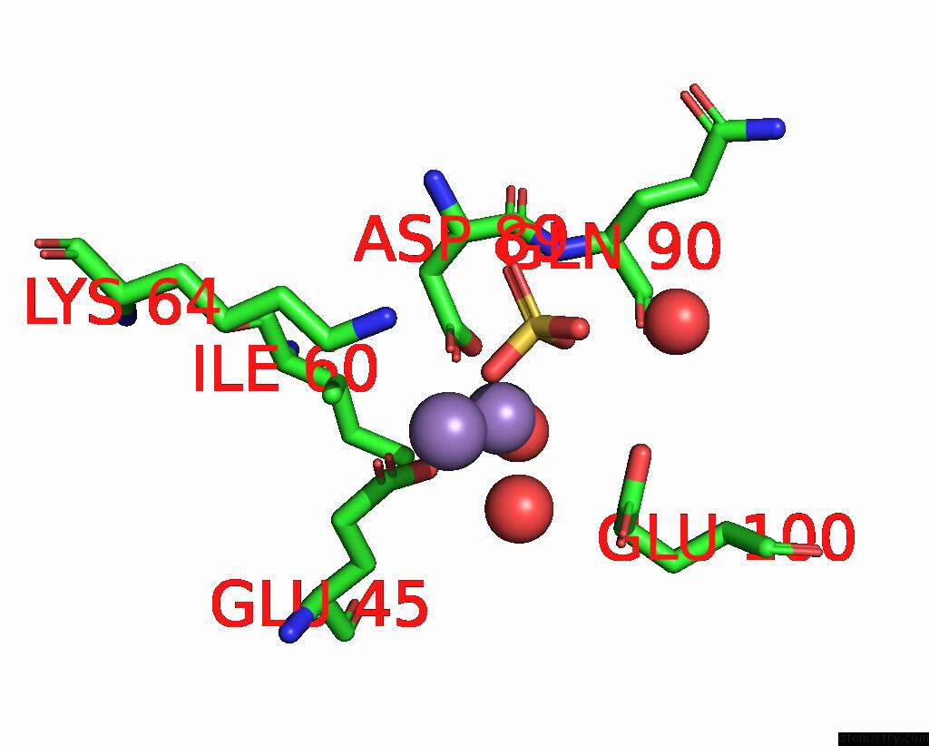
Mono view
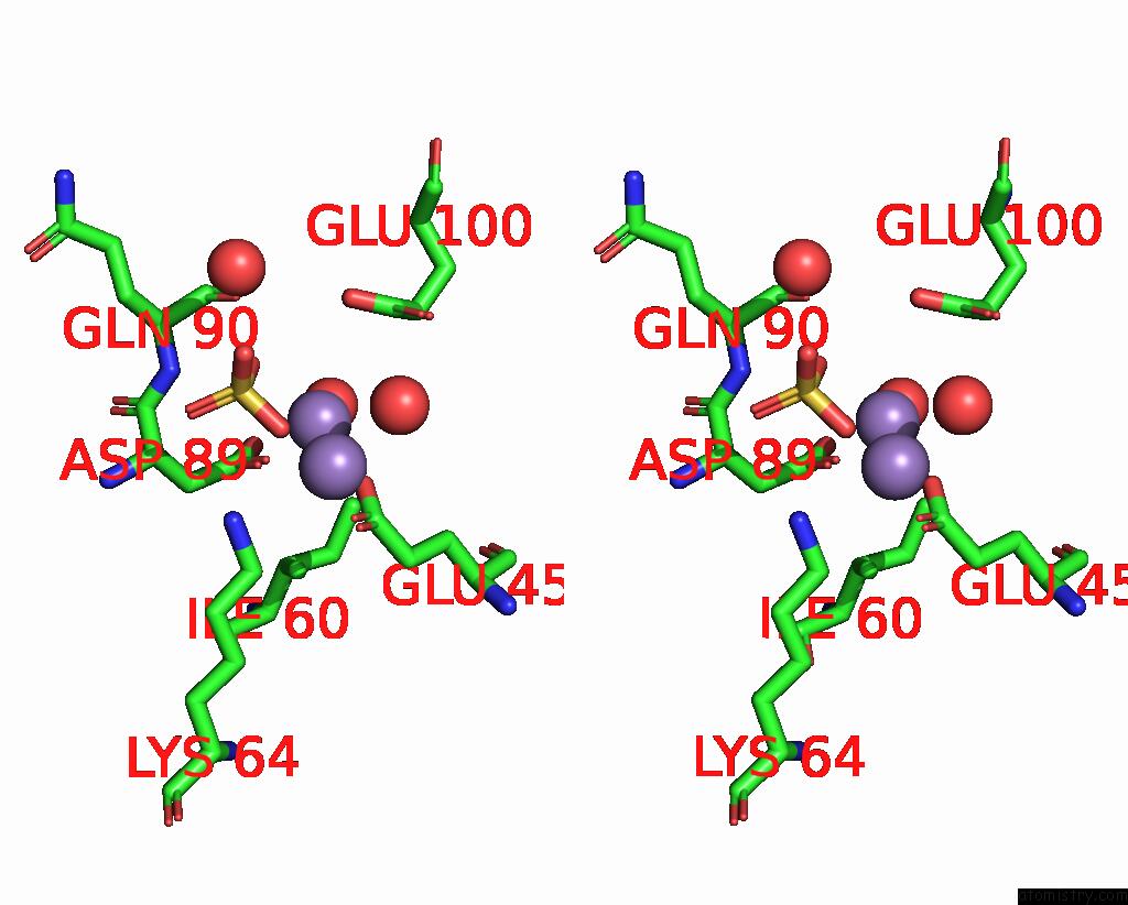
Stereo pair view
Mono view
Stereo pair view
A full contact list of Manganese with other atoms in the Mn binding
site number 2 of Crystal Structure of Maf-Like Protein TBRU21784AAA From T.Brucei within 5.0Å range:
|
Manganese binding site 3 out of 3 in 2amh
Go back to
Manganese binding site 3 out
of 3 in the Crystal Structure of Maf-Like Protein TBRU21784AAA From T.Brucei
Mono view
Stereo pair view
Mono view
Stereo pair view
A full contact list of Manganese with other atoms in the Mn binding
site number 3 of Crystal Structure of Maf-Like Protein TBRU21784AAA From T.Brucei within 5.0Å range:
|
Reference:
J.Caruthers,
E.Merritt,
Structural Genomics Of Pathogenic Protozoa Consortium (Sgpp).
Crystal Structure of Maf-Like Protein TBRU21784AAA From T.Brucei To Be Published.
Page generated: Sat Aug 16 09:44:24 2025
Last articles
Mn in 6JLMMn in 6JLJ
Mn in 6JKV
Mn in 6JKI
Mn in 6J42
Mn in 6IWR
Mn in 6J5Y
Mn in 6J5X
Mn in 6IWQ
Mn in 6IPU