Manganese »
PDB 6i90-6jr1 »
6j5y »
Manganese in PDB 6j5y: Crystal Structure of Fumarylpyruvate Hydrolase From Pseudomonas Aeruginosa in Complex with MN2+ and Pyruvate
Protein crystallography data
The structure of Crystal Structure of Fumarylpyruvate Hydrolase From Pseudomonas Aeruginosa in Complex with MN2+ and Pyruvate, PDB code: 6j5y
was solved by
H.Hong,
H.Seo,
K.-J.Kim,
W.Park,
with X-Ray Crystallography technique. A brief refinement statistics is given in the table below:
| Resolution Low / High (Å) | 32.81 / 1.98 |
| Space group | C 2 2 21 |
| Cell size a, b, c (Å), α, β, γ (°) | 58.090, 61.029, 104.646, 90.00, 90.00, 90.00 |
| R / Rfree (%) | 18.4 / 22.7 |
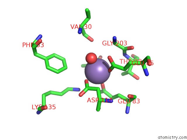
Manganese Binding Sites:
The binding sites of Manganese atom in the Crystal Structure of Fumarylpyruvate Hydrolase From Pseudomonas Aeruginosa in Complex with MN2+ and Pyruvate
(pdb code 6j5y). This binding sites where shown within
5.0 Angstroms radius around Manganese atom.
In total only one binding site of Manganese was determined in the Crystal Structure of Fumarylpyruvate Hydrolase From Pseudomonas Aeruginosa in Complex with MN2+ and Pyruvate, PDB code: 6j5y:
In total only one binding site of Manganese was determined in the Crystal Structure of Fumarylpyruvate Hydrolase From Pseudomonas Aeruginosa in Complex with MN2+ and Pyruvate, PDB code: 6j5y:
Manganese binding site 1 out of 1 in 6j5y
Go back to
Manganese binding site 1 out
of 1 in the Crystal Structure of Fumarylpyruvate Hydrolase From Pseudomonas Aeruginosa in Complex with MN2+ and Pyruvate
Mono view

Stereo pair view
Mono view
Stereo pair view
A full contact list of Manganese with other atoms in the Mn binding
site number 1 of Crystal Structure of Fumarylpyruvate Hydrolase From Pseudomonas Aeruginosa in Complex with MN2+ and Pyruvate within 5.0Å range:
|
Reference:
H.Hong,
H.Seo,
W.Park,
K.-J.Kim.
Sequence, Structure and Function-Based Classification of the Broadly Conserved Fah Superfamily Reveals Two Distinct Fumarylpyruvate Hydrolase Subfamilies. Environ.Microbiol. 2019.
ISSN: ESSN 1462-2920
PubMed: 31657110
DOI: 10.1111/1462-2920.14844
Page generated: Sun Oct 6 04:56:38 2024
ISSN: ESSN 1462-2920
PubMed: 31657110
DOI: 10.1111/1462-2920.14844
Last articles
K in 7M2HK in 7MDJ
K in 7MJQ
K in 7MHR
K in 7MJO
K in 7M0D
K in 7M89
K in 7M83
K in 7M2I
K in 7M7T