Manganese »
PDB 6i90-6jr1 »
6iwq »
Manganese in PDB 6iwq: Crystal Structure of Galnac-T7 with MN2+
Protein crystallography data
The structure of Crystal Structure of Galnac-T7 with MN2+, PDB code: 6iwq
was solved by
C.Yu,
Y.X.Yin,
with X-Ray Crystallography technique. A brief refinement statistics is given in the table below:
| Resolution Low / High (Å) | 49.25 / 2.95 |
| Space group | P 21 21 21 |
| Cell size a, b, c (Å), α, β, γ (°) | 137.695, 158.233, 251.870, 90.00, 90.00, 90.00 |
| R / Rfree (%) | 22.5 / 23.9 |
Manganese Binding Sites:
The binding sites of Manganese atom in the Crystal Structure of Galnac-T7 with MN2+
(pdb code 6iwq). This binding sites where shown within
5.0 Angstroms radius around Manganese atom.
In total 6 binding sites of Manganese where determined in the Crystal Structure of Galnac-T7 with MN2+, PDB code: 6iwq:
Jump to Manganese binding site number: 1; 2; 3; 4; 5; 6;
In total 6 binding sites of Manganese where determined in the Crystal Structure of Galnac-T7 with MN2+, PDB code: 6iwq:
Jump to Manganese binding site number: 1; 2; 3; 4; 5; 6;
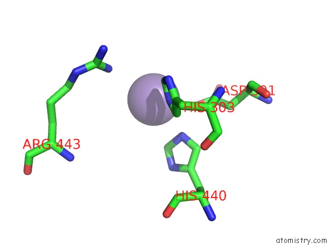
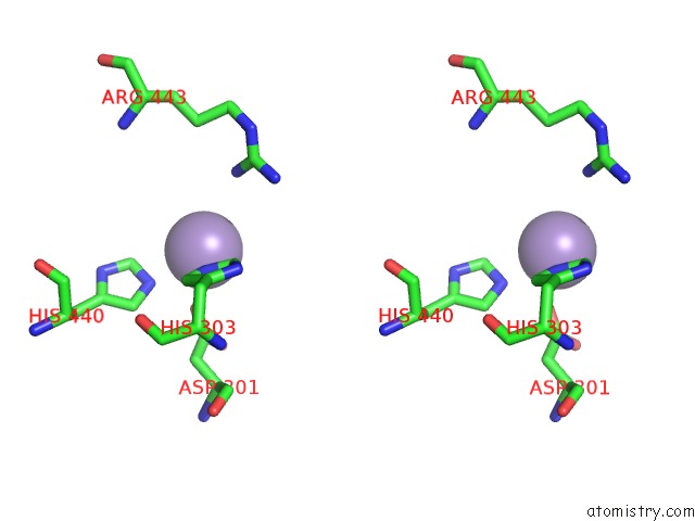
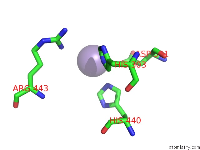
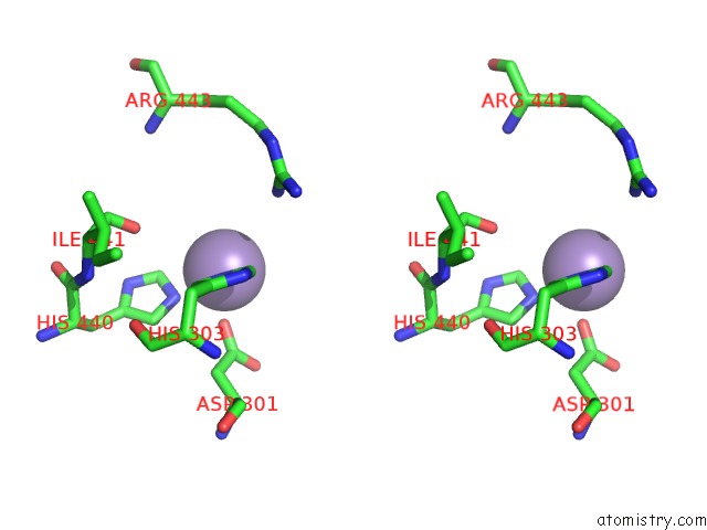
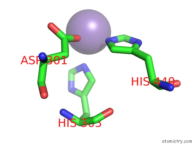
Manganese binding site 1 out of 6 in 6iwq
Go back to
Manganese binding site 1 out
of 6 in the Crystal Structure of Galnac-T7 with MN2+
Mono view
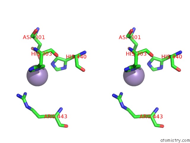
Stereo pair view
Mono view
Stereo pair view
A full contact list of Manganese with other atoms in the Mn binding
site number 1 of Crystal Structure of Galnac-T7 with MN2+ within 5.0Å range:
|
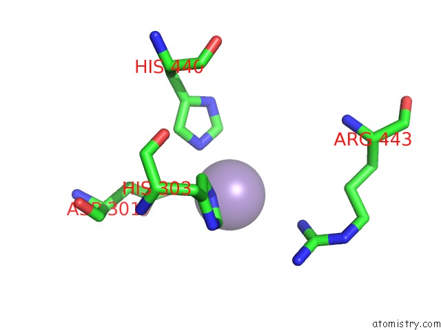
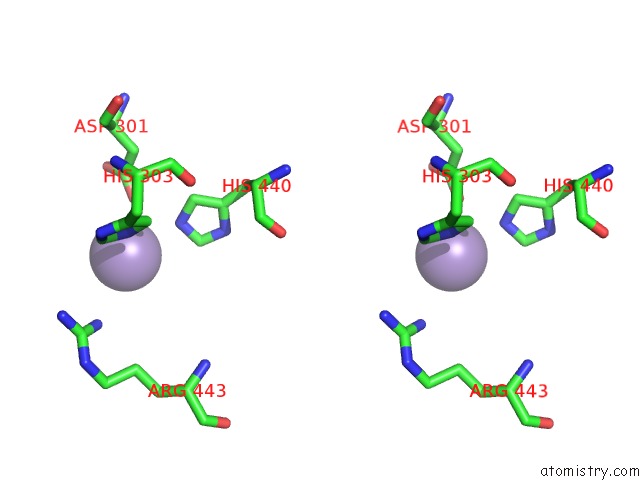
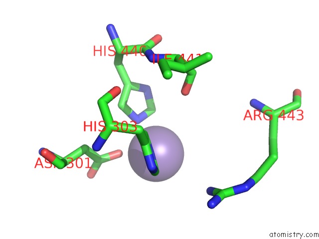
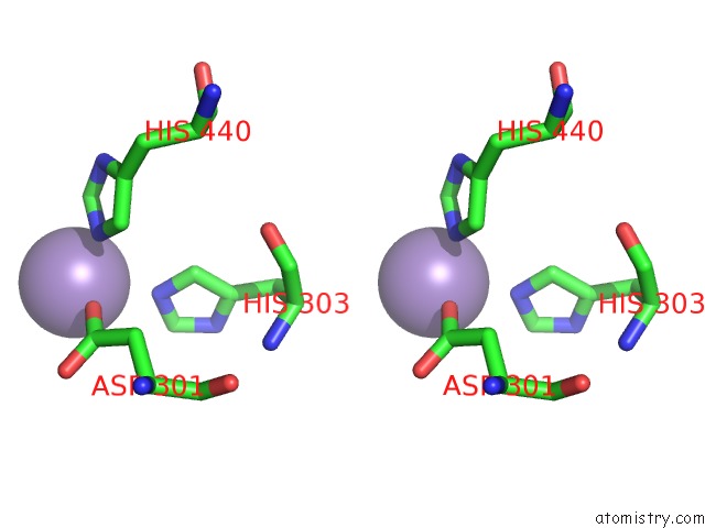
Manganese binding site 2 out of 6 in 6iwq
Go back to
Manganese binding site 2 out
of 6 in the Crystal Structure of Galnac-T7 with MN2+

Mono view
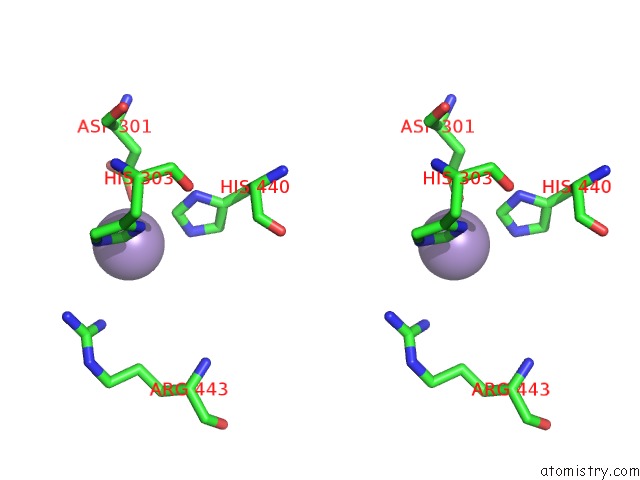
Stereo pair view
Mono view
Stereo pair view
A full contact list of Manganese with other atoms in the Mn binding
site number 2 of Crystal Structure of Galnac-T7 with MN2+ within 5.0Å range:
|
Manganese binding site 3 out of 6 in 6iwq
Go back to
Manganese binding site 3 out
of 6 in the Crystal Structure of Galnac-T7 with MN2+
Mono view
Stereo pair view
Mono view
Stereo pair view
A full contact list of Manganese with other atoms in the Mn binding
site number 3 of Crystal Structure of Galnac-T7 with MN2+ within 5.0Å range:
|
Manganese binding site 4 out of 6 in 6iwq
Go back to
Manganese binding site 4 out
of 6 in the Crystal Structure of Galnac-T7 with MN2+
Mono view
Stereo pair view
Mono view
Stereo pair view
A full contact list of Manganese with other atoms in the Mn binding
site number 4 of Crystal Structure of Galnac-T7 with MN2+ within 5.0Å range:
|
Manganese binding site 5 out of 6 in 6iwq
Go back to
Manganese binding site 5 out
of 6 in the Crystal Structure of Galnac-T7 with MN2+
Mono view
Stereo pair view
Mono view
Stereo pair view
A full contact list of Manganese with other atoms in the Mn binding
site number 5 of Crystal Structure of Galnac-T7 with MN2+ within 5.0Å range:
|
Manganese binding site 6 out of 6 in 6iwq
Go back to
Manganese binding site 6 out
of 6 in the Crystal Structure of Galnac-T7 with MN2+
Mono view
Stereo pair view
Mono view
Stereo pair view
A full contact list of Manganese with other atoms in the Mn binding
site number 6 of Crystal Structure of Galnac-T7 with MN2+ within 5.0Å range:
|
Reference:
C.Yu,
L.Liang,
Y.Yin.
Structural Basis of Carbohydrate Transfer Activity of Udp-Galnac: Polypeptide N-Acetylgalactosaminyltransferase 7. Biochem. Biophys. Res. V. 510 266 2019COMMUN..
ISSN: ESSN 1090-2104
PubMed: 30685086
DOI: 10.1016/J.BBRC.2019.01.084
Page generated: Sun Oct 6 04:56:15 2024
ISSN: ESSN 1090-2104
PubMed: 30685086
DOI: 10.1016/J.BBRC.2019.01.084
Last articles
K in 6DXZK in 6DVN
K in 6DVM
K in 6DWO
K in 6DXV
K in 6DVX
K in 6DVL
K in 6DVO
K in 6DUR
K in 6DPN