Manganese »
PDB 2a7a-2axt »
2am4 »
Manganese in PDB 2am4: Crystal Structure of N-Acetylglucosaminyltransferase I in Complex with Udp-2-Deoxy-2-Fluoro-Glucose
Enzymatic activity of Crystal Structure of N-Acetylglucosaminyltransferase I in Complex with Udp-2-Deoxy-2-Fluoro-Glucose
All present enzymatic activity of Crystal Structure of N-Acetylglucosaminyltransferase I in Complex with Udp-2-Deoxy-2-Fluoro-Glucose:
2.4.1.101;
2.4.1.101;
Protein crystallography data
The structure of Crystal Structure of N-Acetylglucosaminyltransferase I in Complex with Udp-2-Deoxy-2-Fluoro-Glucose, PDB code: 2am4
was solved by
J.M.Rini,
R.D.Gordon,
with X-Ray Crystallography technique. A brief refinement statistics is given in the table below:
| Resolution Low / High (Å) | 29.80 / 1.70 |
| Space group | P 21 21 21 |
| Cell size a, b, c (Å), α, β, γ (°) | 40.917, 82.501, 102.394, 90.00, 90.00, 90.00 |
| R / Rfree (%) | 19.3 / 21.7 |
Other elements in 2am4:
The structure of Crystal Structure of N-Acetylglucosaminyltransferase I in Complex with Udp-2-Deoxy-2-Fluoro-Glucose also contains other interesting chemical elements:
| Fluorine | (F) | 1 atom |
Manganese Binding Sites:
The binding sites of Manganese atom in the Crystal Structure of N-Acetylglucosaminyltransferase I in Complex with Udp-2-Deoxy-2-Fluoro-Glucose
(pdb code 2am4). This binding sites where shown within
5.0 Angstroms radius around Manganese atom.
In total only one binding site of Manganese was determined in the Crystal Structure of N-Acetylglucosaminyltransferase I in Complex with Udp-2-Deoxy-2-Fluoro-Glucose, PDB code: 2am4:
In total only one binding site of Manganese was determined in the Crystal Structure of N-Acetylglucosaminyltransferase I in Complex with Udp-2-Deoxy-2-Fluoro-Glucose, PDB code: 2am4:
Manganese binding site 1 out of 1 in 2am4
Go back to
Manganese binding site 1 out
of 1 in the Crystal Structure of N-Acetylglucosaminyltransferase I in Complex with Udp-2-Deoxy-2-Fluoro-Glucose
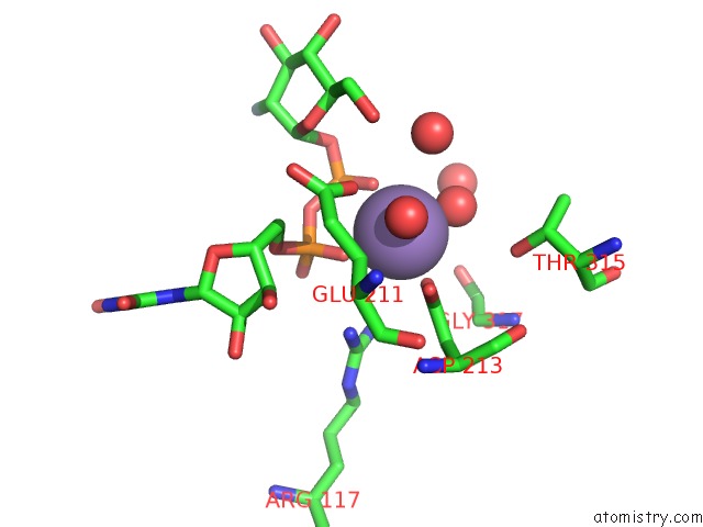
Mono view
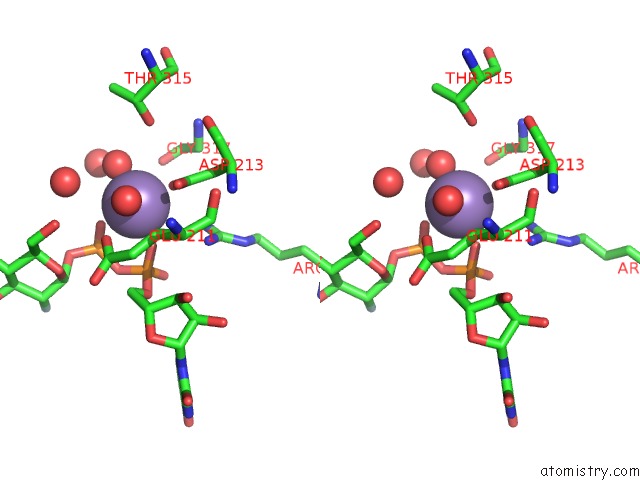
Stereo pair view
Mono view
Stereo pair view
A full contact list of Manganese with other atoms in the Mn binding
site number 1 of Crystal Structure of N-Acetylglucosaminyltransferase I in Complex with Udp-2-Deoxy-2-Fluoro-Glucose within 5.0Å range:
|
Reference:
R.D.Gordon,
P.Sivarajah,
M.Satkunarajah,
D.Ma,
C.A.Tarling,
D.Vizitiu,
S.G.Withers,
J.M.Rini.
X-Ray Crystal Structures of Rabbit N-Acetylglucosaminyltransferase I (Gnt I) in Complex with Donor Substrate Analogues. J.Mol.Biol. V. 360 67 2006.
ISSN: ISSN 0022-2836
PubMed: 16769084
DOI: 10.1016/J.JMB.2006.04.058
Page generated: Sat Aug 16 09:44:21 2025
ISSN: ISSN 0022-2836
PubMed: 16769084
DOI: 10.1016/J.JMB.2006.04.058
Last articles
Mn in 6JLMMn in 6JLJ
Mn in 6JKV
Mn in 6JKI
Mn in 6J42
Mn in 6IWR
Mn in 6J5Y
Mn in 6J5X
Mn in 6IWQ
Mn in 6IPU