Manganese »
PDB 8zge-9bya »
8zwd »
Manganese in PDB 8zwd: Crystal Structure of Methanol DEHYDROGENASE1 From Bacillus Methanolicus
Enzymatic activity of Crystal Structure of Methanol DEHYDROGENASE1 From Bacillus Methanolicus
All present enzymatic activity of Crystal Structure of Methanol DEHYDROGENASE1 From Bacillus Methanolicus:
1.1.1.244;
1.1.1.244;
Protein crystallography data
The structure of Crystal Structure of Methanol DEHYDROGENASE1 From Bacillus Methanolicus, PDB code: 8zwd
was solved by
B.D.Ma,
X.D.Kong,
with X-Ray Crystallography technique. A brief refinement statistics is given in the table below:
| Resolution Low / High (Å) | 29.78 / 3.00 |
| Space group | P 21 21 21 |
| Cell size a, b, c (Å), α, β, γ (°) | 256.022, 83.578, 220.557, 90, 90, 90 |
| R / Rfree (%) | 21.3 / 26 |
Other elements in 8zwd:
The structure of Crystal Structure of Methanol DEHYDROGENASE1 From Bacillus Methanolicus also contains other interesting chemical elements:
| Chlorine | (Cl) | 2 atoms |
Manganese Binding Sites:
The binding sites of Manganese atom in the Crystal Structure of Methanol DEHYDROGENASE1 From Bacillus Methanolicus
(pdb code 8zwd). This binding sites where shown within
5.0 Angstroms radius around Manganese atom.
In total 10 binding sites of Manganese where determined in the Crystal Structure of Methanol DEHYDROGENASE1 From Bacillus Methanolicus, PDB code: 8zwd:
Jump to Manganese binding site number: 1; 2; 3; 4; 5; 6; 7; 8; 9; 10;
In total 10 binding sites of Manganese where determined in the Crystal Structure of Methanol DEHYDROGENASE1 From Bacillus Methanolicus, PDB code: 8zwd:
Jump to Manganese binding site number: 1; 2; 3; 4; 5; 6; 7; 8; 9; 10;
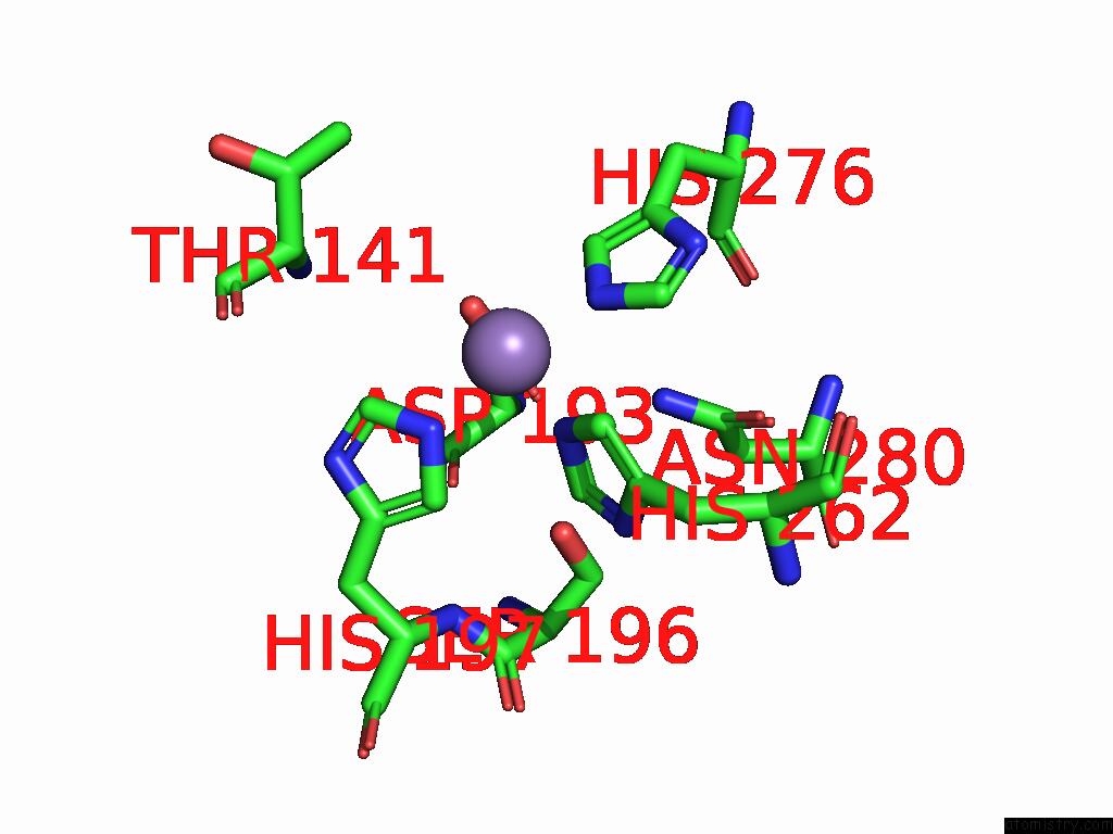
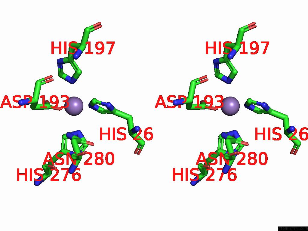
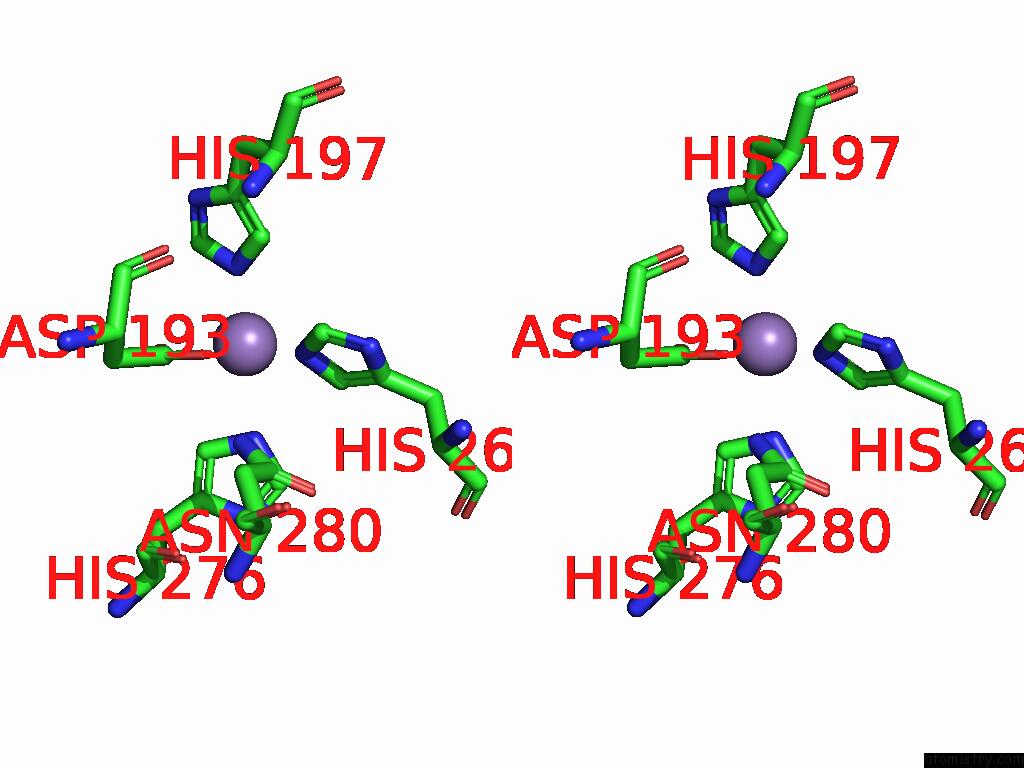
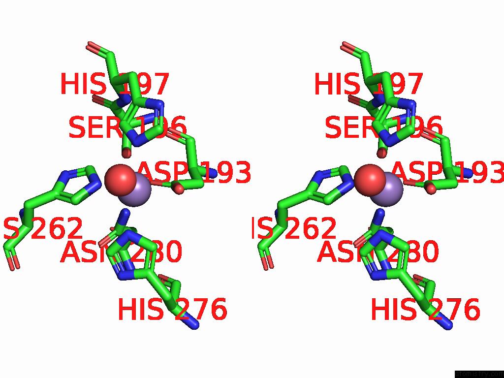
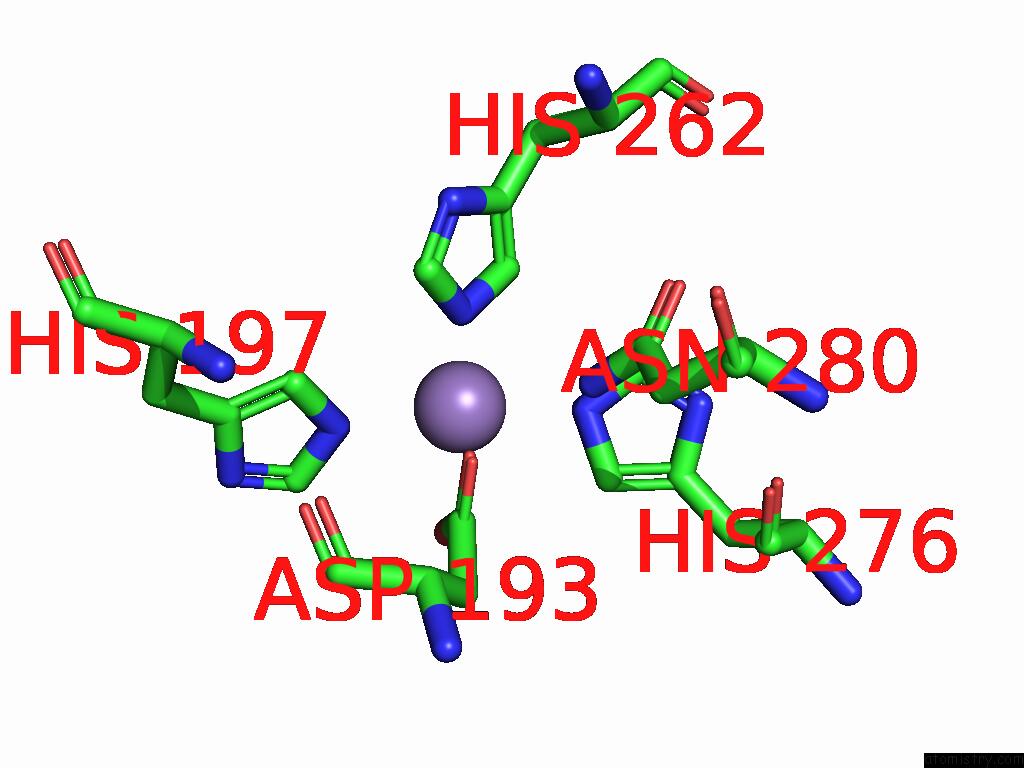
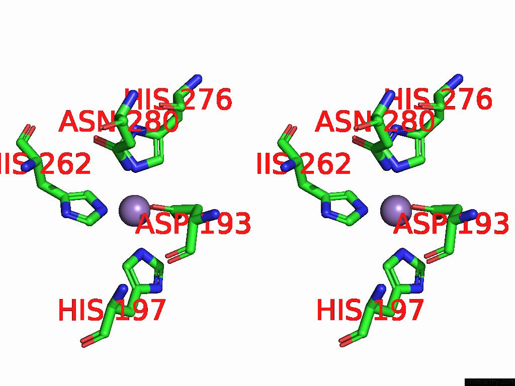
Manganese binding site 1 out of 10 in 8zwd
Go back to
Manganese binding site 1 out
of 10 in the Crystal Structure of Methanol DEHYDROGENASE1 From Bacillus Methanolicus
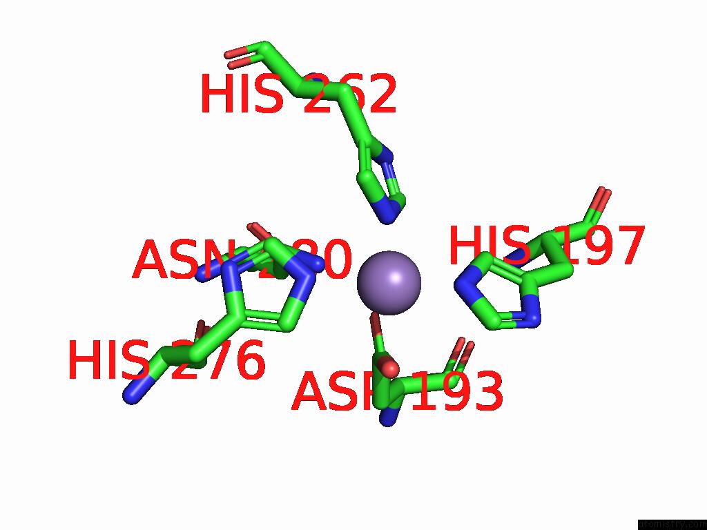
Mono view
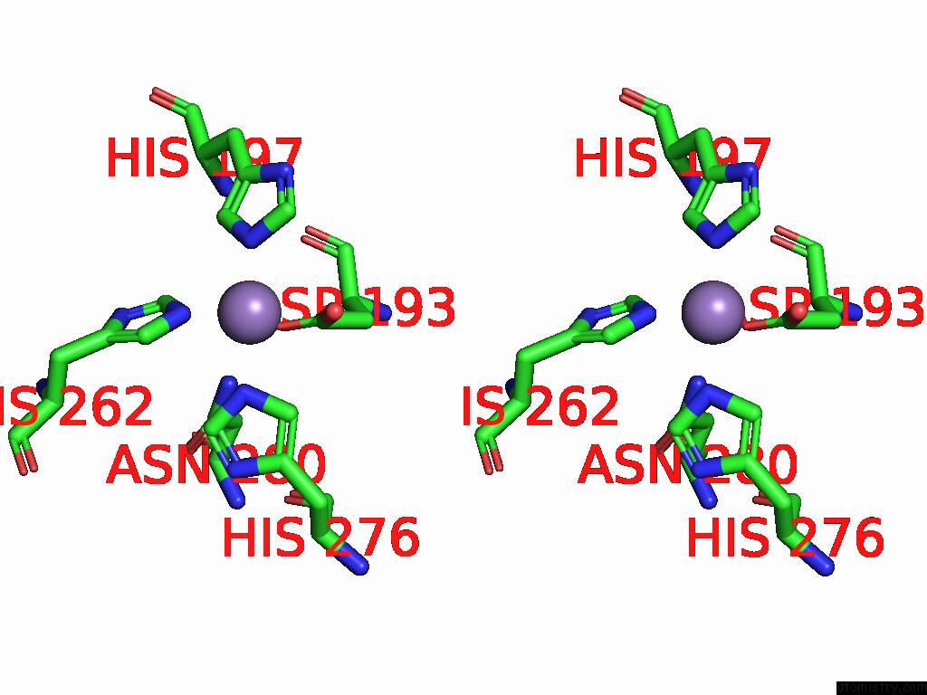
Stereo pair view
Mono view
Stereo pair view
A full contact list of Manganese with other atoms in the Mn binding
site number 1 of Crystal Structure of Methanol DEHYDROGENASE1 From Bacillus Methanolicus within 5.0Å range:
|
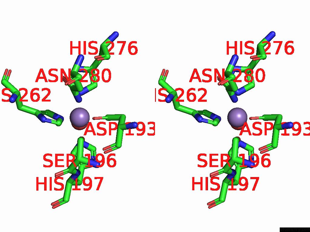
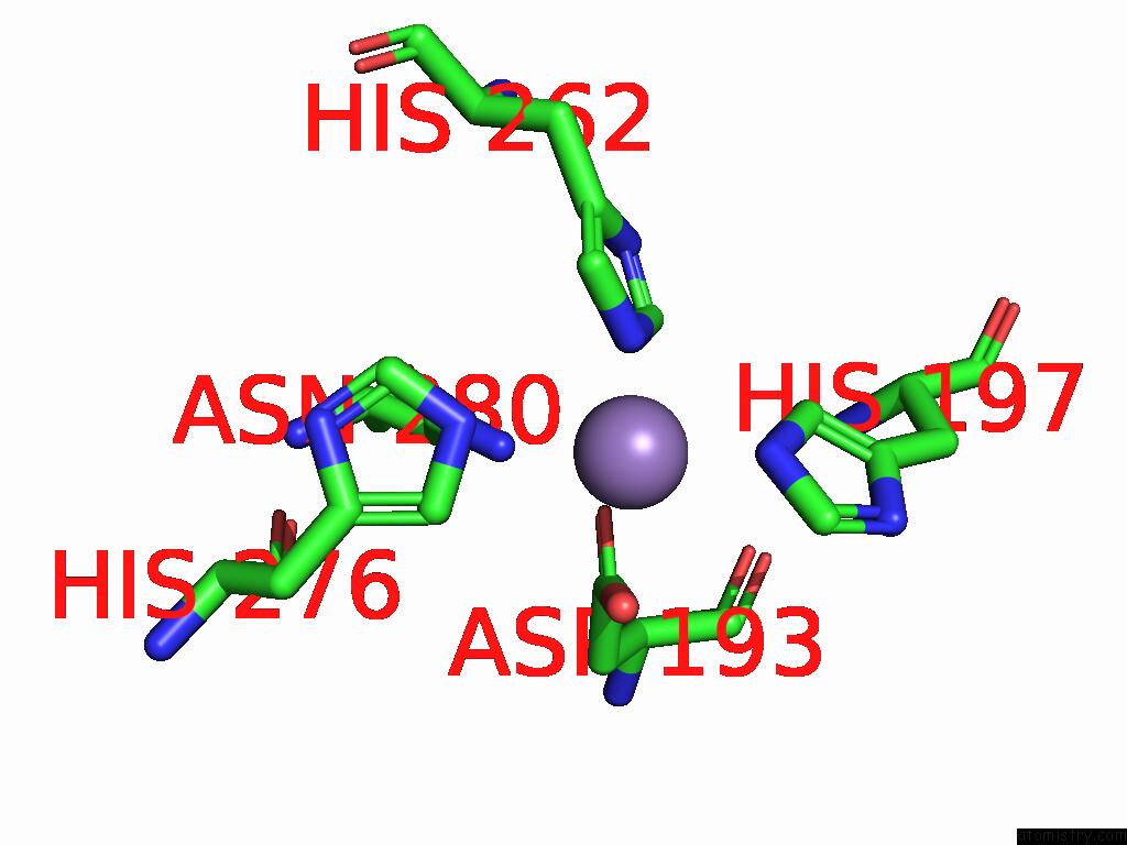
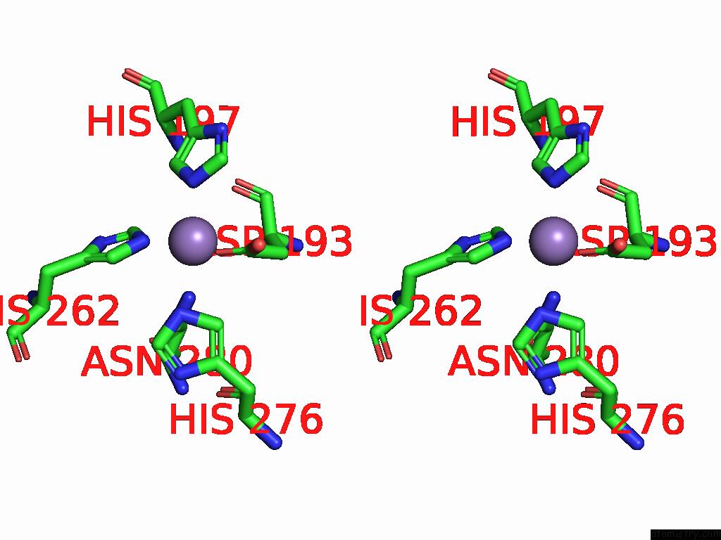
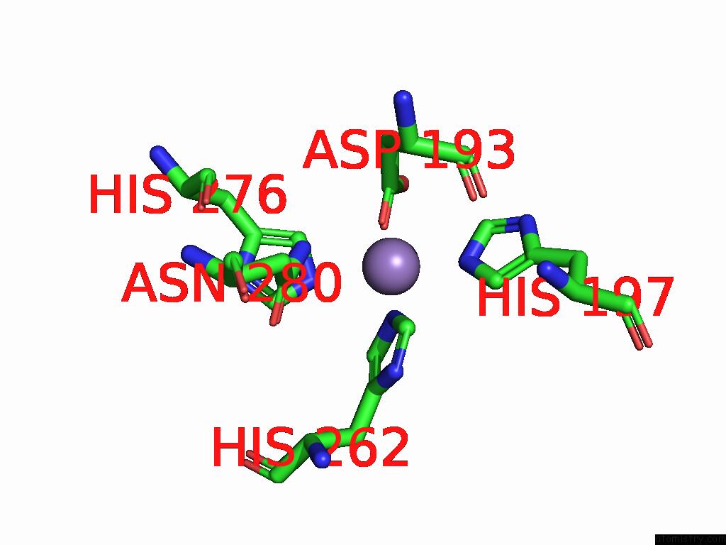
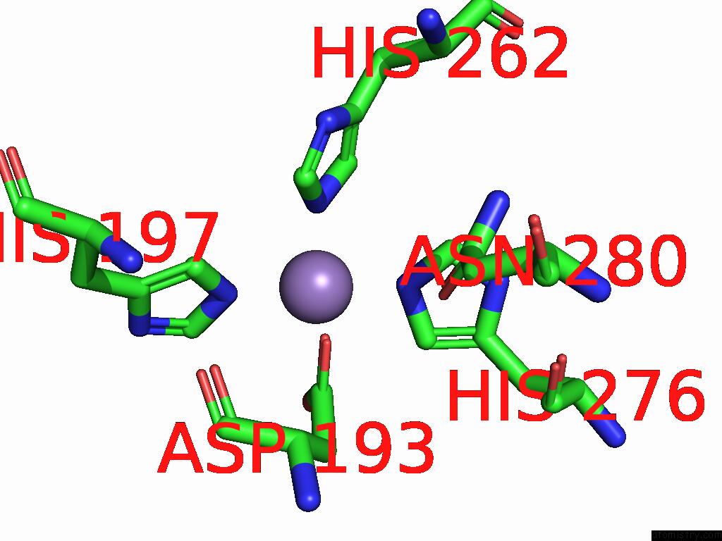
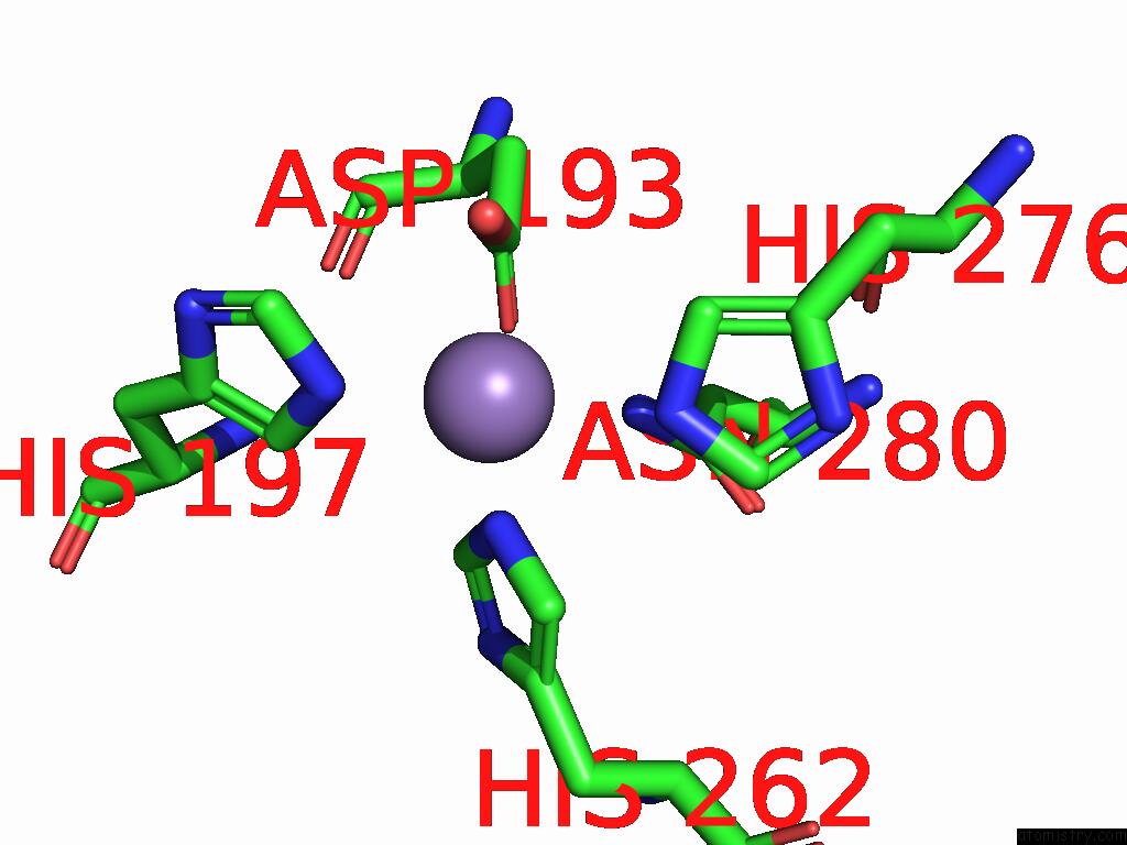
Manganese binding site 2 out of 10 in 8zwd
Go back to
Manganese binding site 2 out
of 10 in the Crystal Structure of Methanol DEHYDROGENASE1 From Bacillus Methanolicus
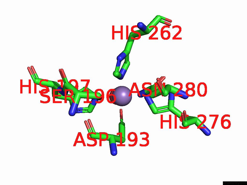
Mono view
Stereo pair view
Mono view
Stereo pair view
A full contact list of Manganese with other atoms in the Mn binding
site number 2 of Crystal Structure of Methanol DEHYDROGENASE1 From Bacillus Methanolicus within 5.0Å range:
|
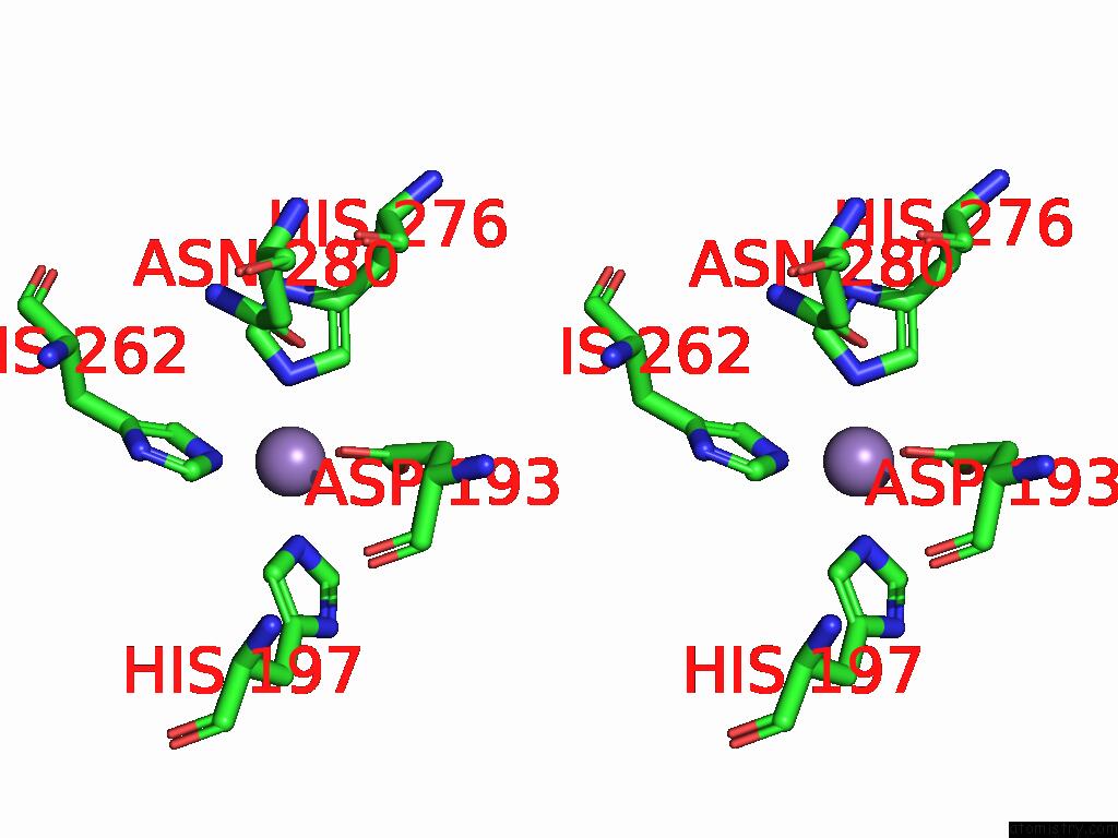
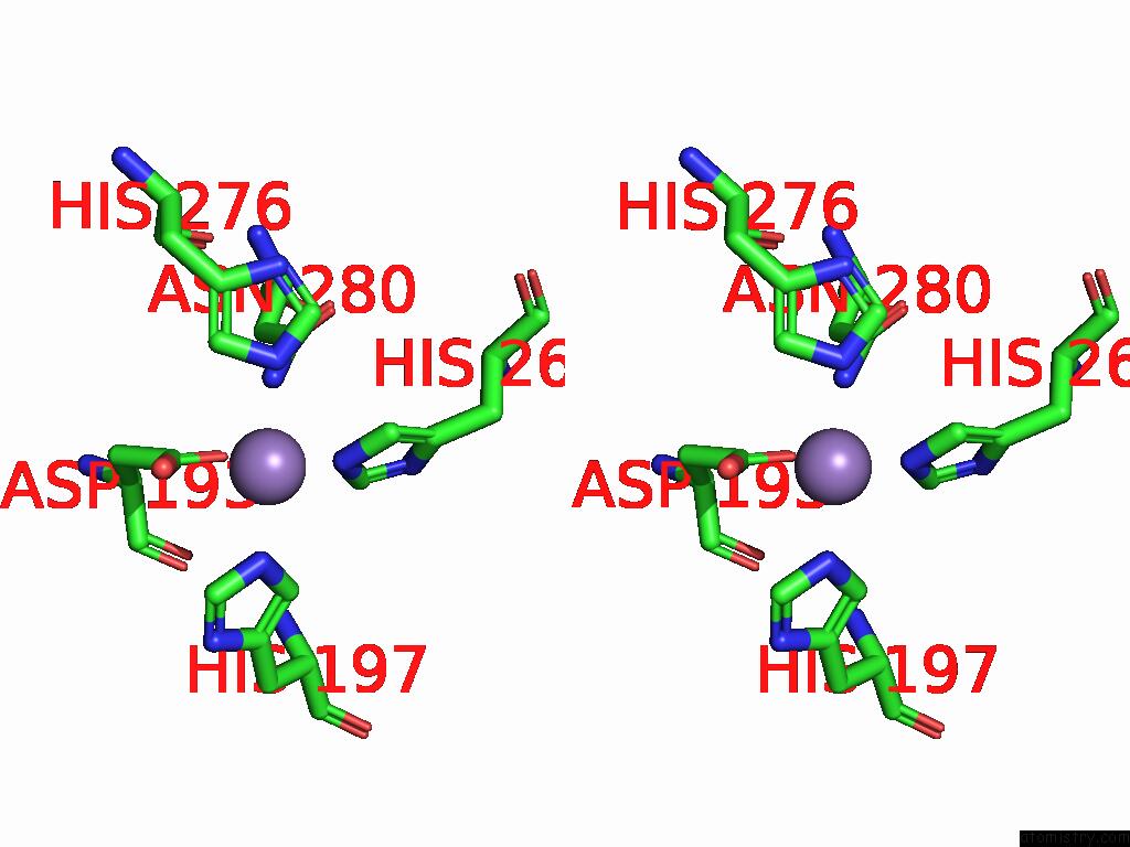
Manganese binding site 3 out of 10 in 8zwd
Go back to
Manganese binding site 3 out
of 10 in the Crystal Structure of Methanol DEHYDROGENASE1 From Bacillus Methanolicus
Mono view

Stereo pair view
Mono view
Stereo pair view
A full contact list of Manganese with other atoms in the Mn binding
site number 3 of Crystal Structure of Methanol DEHYDROGENASE1 From Bacillus Methanolicus within 5.0Å range:
|
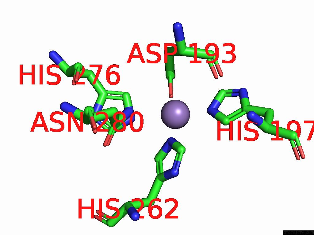
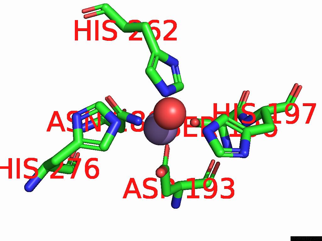
Manganese binding site 4 out of 10 in 8zwd
Go back to
Manganese binding site 4 out
of 10 in the Crystal Structure of Methanol DEHYDROGENASE1 From Bacillus Methanolicus
Mono view
Stereo pair view
Mono view
Stereo pair view
A full contact list of Manganese with other atoms in the Mn binding
site number 4 of Crystal Structure of Methanol DEHYDROGENASE1 From Bacillus Methanolicus within 5.0Å range:
|
Manganese binding site 5 out of 10 in 8zwd
Go back to
Manganese binding site 5 out
of 10 in the Crystal Structure of Methanol DEHYDROGENASE1 From Bacillus Methanolicus
Mono view
Stereo pair view
Mono view
Stereo pair view
A full contact list of Manganese with other atoms in the Mn binding
site number 5 of Crystal Structure of Methanol DEHYDROGENASE1 From Bacillus Methanolicus within 5.0Å range:
|
Manganese binding site 6 out of 10 in 8zwd
Go back to
Manganese binding site 6 out
of 10 in the Crystal Structure of Methanol DEHYDROGENASE1 From Bacillus Methanolicus
Mono view
Stereo pair view
Mono view
Stereo pair view
A full contact list of Manganese with other atoms in the Mn binding
site number 6 of Crystal Structure of Methanol DEHYDROGENASE1 From Bacillus Methanolicus within 5.0Å range:
|
Manganese binding site 7 out of 10 in 8zwd
Go back to
Manganese binding site 7 out
of 10 in the Crystal Structure of Methanol DEHYDROGENASE1 From Bacillus Methanolicus
Mono view
Stereo pair view
Mono view
Stereo pair view
A full contact list of Manganese with other atoms in the Mn binding
site number 7 of Crystal Structure of Methanol DEHYDROGENASE1 From Bacillus Methanolicus within 5.0Å range:
|
Manganese binding site 8 out of 10 in 8zwd
Go back to
Manganese binding site 8 out
of 10 in the Crystal Structure of Methanol DEHYDROGENASE1 From Bacillus Methanolicus
Mono view
Stereo pair view
Mono view
Stereo pair view
A full contact list of Manganese with other atoms in the Mn binding
site number 8 of Crystal Structure of Methanol DEHYDROGENASE1 From Bacillus Methanolicus within 5.0Å range:
|
Manganese binding site 9 out of 10 in 8zwd
Go back to
Manganese binding site 9 out
of 10 in the Crystal Structure of Methanol DEHYDROGENASE1 From Bacillus Methanolicus
Mono view
Stereo pair view
Mono view
Stereo pair view
A full contact list of Manganese with other atoms in the Mn binding
site number 9 of Crystal Structure of Methanol DEHYDROGENASE1 From Bacillus Methanolicus within 5.0Å range:
|
Manganese binding site 10 out of 10 in 8zwd
Go back to
Manganese binding site 10 out
of 10 in the Crystal Structure of Methanol DEHYDROGENASE1 From Bacillus Methanolicus
Mono view
Stereo pair view
Mono view
Stereo pair view
A full contact list of Manganese with other atoms in the Mn binding
site number 10 of Crystal Structure of Methanol DEHYDROGENASE1 From Bacillus Methanolicus within 5.0Å range:
|
Reference:
B.D.Ma,
X.D.Kong.
Crystal Structure of Methanol DEHYDROGENASE1 From Bacillus Methanolicus To Be Published.
Page generated: Sun Aug 17 02:06:54 2025
Last articles
Na in 2WGMNa in 2WM2
Na in 2WOF
Na in 2WO0
Na in 2WNT
Na in 2WNH
Na in 2WNN
Na in 2WL4
Na in 2WMZ
Na in 2WLU