Manganese »
PDB 7ewx-7fq9 »
7ex3 »
Manganese in PDB 7ex3: Crystal Structure of Ebinur Lake Virus Cap Snatching Endonuclease in Complex with Inhibitor 3
Enzymatic activity of Crystal Structure of Ebinur Lake Virus Cap Snatching Endonuclease in Complex with Inhibitor 3
All present enzymatic activity of Crystal Structure of Ebinur Lake Virus Cap Snatching Endonuclease in Complex with Inhibitor 3:
2.7.7.48;
2.7.7.48;
Protein crystallography data
The structure of Crystal Structure of Ebinur Lake Virus Cap Snatching Endonuclease in Complex with Inhibitor 3, PDB code: 7ex3
was solved by
W.Kuang,
Z.Hu,
P.Gong,
with X-Ray Crystallography technique. A brief refinement statistics is given in the table below:
| Resolution Low / High (Å) | 35.63 / 2.25 |
| Space group | P 21 21 21 |
| Cell size a, b, c (Å), α, β, γ (°) | 40.666, 70.873, 71.253, 90, 90, 90 |
| R / Rfree (%) | 19 / 22.6 |
Other elements in 7ex3:
The structure of Crystal Structure of Ebinur Lake Virus Cap Snatching Endonuclease in Complex with Inhibitor 3 also contains other interesting chemical elements:
| Chlorine | (Cl) | 4 atoms |
| Fluorine | (F) | 1 atom |
Manganese Binding Sites:
The binding sites of Manganese atom in the Crystal Structure of Ebinur Lake Virus Cap Snatching Endonuclease in Complex with Inhibitor 3
(pdb code 7ex3). This binding sites where shown within
5.0 Angstroms radius around Manganese atom.
In total 2 binding sites of Manganese where determined in the Crystal Structure of Ebinur Lake Virus Cap Snatching Endonuclease in Complex with Inhibitor 3, PDB code: 7ex3:
Jump to Manganese binding site number: 1; 2;
In total 2 binding sites of Manganese where determined in the Crystal Structure of Ebinur Lake Virus Cap Snatching Endonuclease in Complex with Inhibitor 3, PDB code: 7ex3:
Jump to Manganese binding site number: 1; 2;
Manganese binding site 1 out of 2 in 7ex3
Go back to
Manganese binding site 1 out
of 2 in the Crystal Structure of Ebinur Lake Virus Cap Snatching Endonuclease in Complex with Inhibitor 3
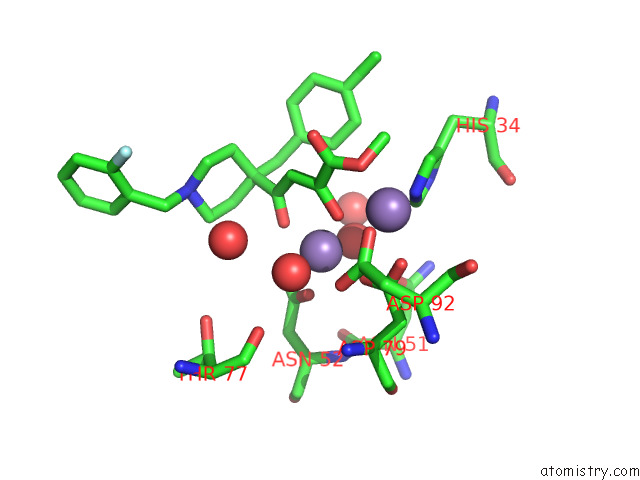
Mono view
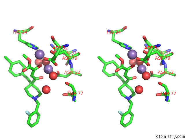
Stereo pair view
Mono view
Stereo pair view
A full contact list of Manganese with other atoms in the Mn binding
site number 1 of Crystal Structure of Ebinur Lake Virus Cap Snatching Endonuclease in Complex with Inhibitor 3 within 5.0Å range:
|
Manganese binding site 2 out of 2 in 7ex3
Go back to
Manganese binding site 2 out
of 2 in the Crystal Structure of Ebinur Lake Virus Cap Snatching Endonuclease in Complex with Inhibitor 3
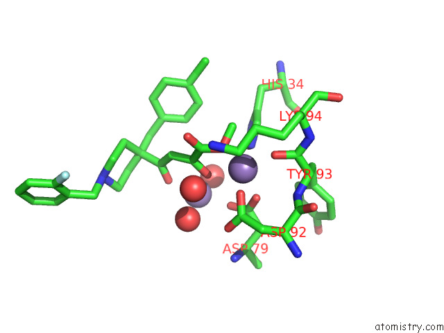
Mono view
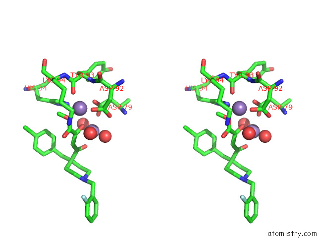
Stereo pair view
Mono view
Stereo pair view
A full contact list of Manganese with other atoms in the Mn binding
site number 2 of Crystal Structure of Ebinur Lake Virus Cap Snatching Endonuclease in Complex with Inhibitor 3 within 5.0Å range:
|
Reference:
W.Kuang,
H.Zhang,
Y.Cai,
G.Zhang,
F.Deng,
H.Li,
Y.Zhou,
M.Wang,
P.Gong,
Y.Guo,
Z.Hu.
Structural and Biochemical Basis For Development of Diketo Acid Inhibitors Targeting the Cap-Snatching Endonuclease of the Ebinur Lake Virus (Order: Bunyavirales ). J.Virol. V. 96 17321 2022.
ISSN: ESSN 1098-5514
PubMed: 35266805
DOI: 10.1128/JVI.02173-21
Page generated: Sun Oct 6 08:37:02 2024
ISSN: ESSN 1098-5514
PubMed: 35266805
DOI: 10.1128/JVI.02173-21
Last articles
K in 2B1GK in 2B1I
K in 2ATK
K in 2AVH
K in 2ATS
K in 2APO
K in 2AS4
K in 2ASC
K in 2AS3
K in 2AOP