Manganese »
PDB 5jqk-5kg4 »
5jqk »
Manganese in PDB 5jqk: The Xray Crystal Structure of P. Falciparum Aminopeptidase P
Protein crystallography data
The structure of The Xray Crystal Structure of P. Falciparum Aminopeptidase P, PDB code: 5jqk
was solved by
N.Drinkwater,
S.Mcgowan,
with X-Ray Crystallography technique. A brief refinement statistics is given in the table below:
| Resolution Low / High (Å) | 44.99 / 2.35 |
| Space group | C 1 2 1 |
| Cell size a, b, c (Å), α, β, γ (°) | 146.740, 100.070, 106.660, 90.00, 105.37, 90.00 |
| R / Rfree (%) | 18.9 / 22.6 |
Manganese Binding Sites:
The binding sites of Manganese atom in the The Xray Crystal Structure of P. Falciparum Aminopeptidase P
(pdb code 5jqk). This binding sites where shown within
5.0 Angstroms radius around Manganese atom.
In total 4 binding sites of Manganese where determined in the The Xray Crystal Structure of P. Falciparum Aminopeptidase P, PDB code: 5jqk:
Jump to Manganese binding site number: 1; 2; 3; 4;
In total 4 binding sites of Manganese where determined in the The Xray Crystal Structure of P. Falciparum Aminopeptidase P, PDB code: 5jqk:
Jump to Manganese binding site number: 1; 2; 3; 4;
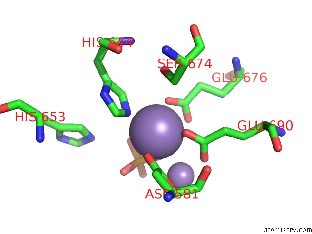
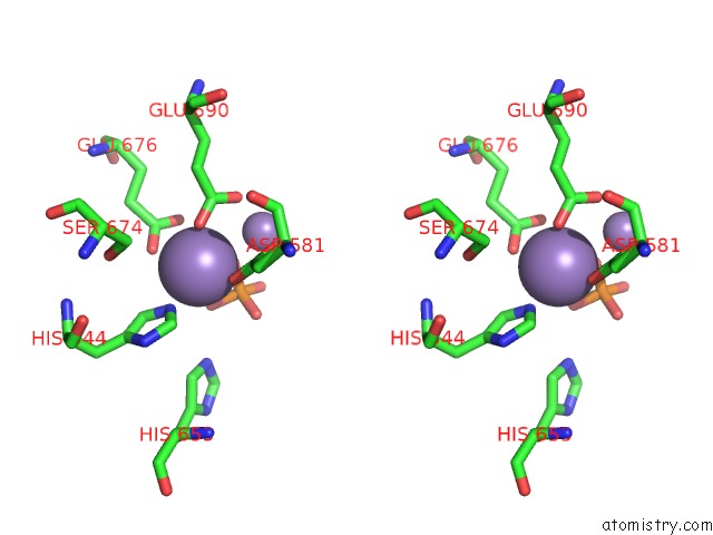
Manganese binding site 1 out of 4 in 5jqk
Go back to
Manganese binding site 1 out
of 4 in the The Xray Crystal Structure of P. Falciparum Aminopeptidase P
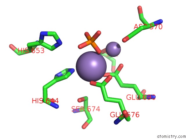
Mono view
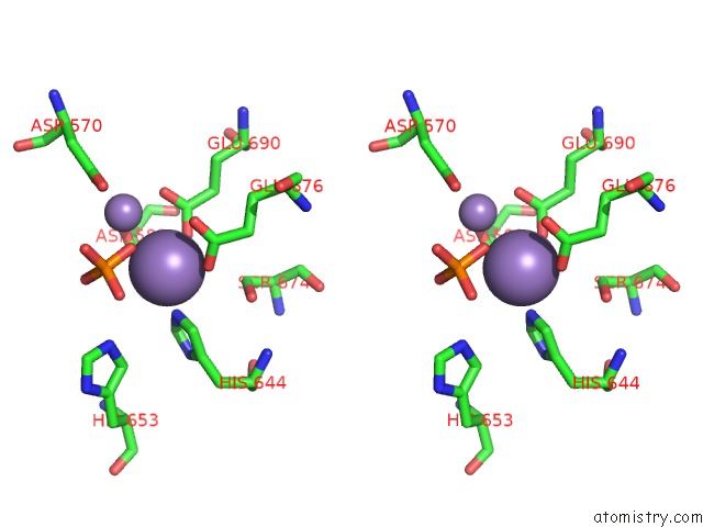
Stereo pair view
Mono view
Stereo pair view
A full contact list of Manganese with other atoms in the Mn binding
site number 1 of The Xray Crystal Structure of P. Falciparum Aminopeptidase P within 5.0Å range:
|
Manganese binding site 2 out of 4 in 5jqk
Go back to
Manganese binding site 2 out
of 4 in the The Xray Crystal Structure of P. Falciparum Aminopeptidase P
Mono view
Stereo pair view
Mono view
Stereo pair view
A full contact list of Manganese with other atoms in the Mn binding
site number 2 of The Xray Crystal Structure of P. Falciparum Aminopeptidase P within 5.0Å range:
|
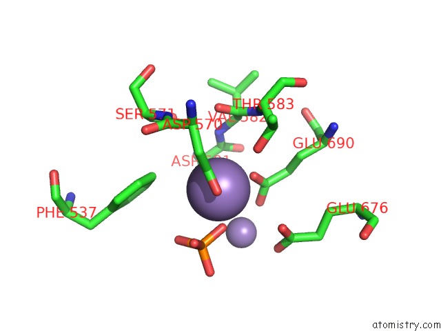
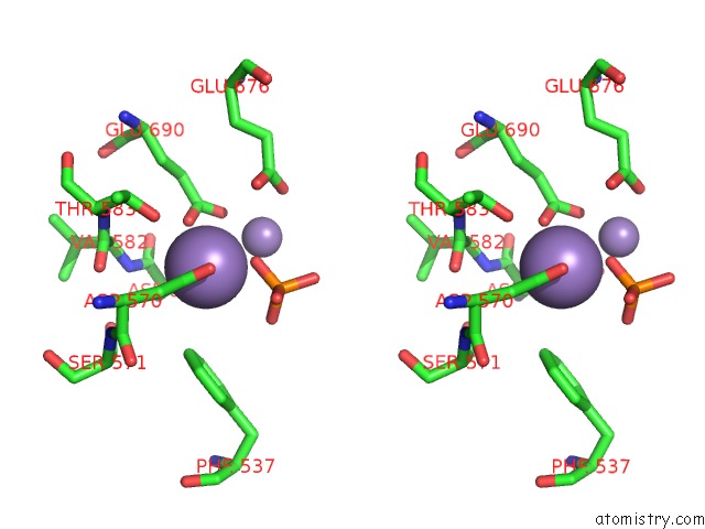
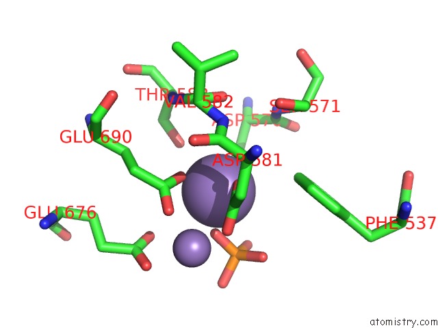
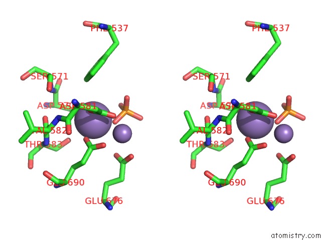
Manganese binding site 3 out of 4 in 5jqk
Go back to
Manganese binding site 3 out
of 4 in the The Xray Crystal Structure of P. Falciparum Aminopeptidase P
Mono view
Stereo pair view
Mono view
Stereo pair view
A full contact list of Manganese with other atoms in the Mn binding
site number 3 of The Xray Crystal Structure of P. Falciparum Aminopeptidase P within 5.0Å range:
|
Manganese binding site 4 out of 4 in 5jqk
Go back to
Manganese binding site 4 out
of 4 in the The Xray Crystal Structure of P. Falciparum Aminopeptidase P
Mono view
Stereo pair view
Mono view
Stereo pair view
A full contact list of Manganese with other atoms in the Mn binding
site number 4 of The Xray Crystal Structure of P. Falciparum Aminopeptidase P within 5.0Å range:
|
Reference:
N.Drinkwater,
K.K.Sivaraman,
R.S.Bamert,
W.Rut,
K.Mohamed,
N.B.Vinh,
P.J.Scammells,
M.Drag,
S.Mcgowan.
Structure and Substrate Fingerprint of Aminopeptidase P From Plasmodium Falciparum. Biochem.J. V. 473 3189 2016.
ISSN: ESSN 1470-8728
PubMed: 27462122
DOI: 10.1042/BCJ20160550
Page generated: Sun Oct 6 01:34:39 2024
ISSN: ESSN 1470-8728
PubMed: 27462122
DOI: 10.1042/BCJ20160550
Last articles
Mg in 1KZUMg in 1L2X
Mg in 1L3P
Mg in 1L3J
Mg in 1L2O
Mg in 1L2E
Mg in 1L0O
Mg in 1L1R
Mg in 1KXG
Mg in 1KYR