Manganese »
PDB 5cdn-5dcb »
5cna »
Manganese in PDB 5cna: Refined Structure of Concanavalin A Complexed with Alpha- Methyl-D-Mannopyranoside at 2.0 Angstroms Resolution and Comparison with the Saccharide-Free Structure
Protein crystallography data
The structure of Refined Structure of Concanavalin A Complexed with Alpha- Methyl-D-Mannopyranoside at 2.0 Angstroms Resolution and Comparison with the Saccharide-Free Structure, PDB code: 5cna
was solved by
J.H.Naismith,
C.Emmerich,
J.Habash,
S.J.Harrop,
J.R.Helliwell,
W.N.Hunter,
J.Raftery,
A.J.Kalb(Gilboa),
J.Yariv,
with X-Ray Crystallography technique. A brief refinement statistics is given in the table below:
| Resolution Low / High (Å) | 8.00 / 2.00 |
| Space group | P 21 21 21 |
| Cell size a, b, c (Å), α, β, γ (°) | 123.700, 128.600, 67.200, 90.00, 90.00, 90.00 |
| R / Rfree (%) | 19.9 / n/a |
Other elements in 5cna:
The structure of Refined Structure of Concanavalin A Complexed with Alpha- Methyl-D-Mannopyranoside at 2.0 Angstroms Resolution and Comparison with the Saccharide-Free Structure also contains other interesting chemical elements:
| Chlorine | (Cl) | 1 atom |
| Calcium | (Ca) | 4 atoms |
Manganese Binding Sites:
The binding sites of Manganese atom in the Refined Structure of Concanavalin A Complexed with Alpha- Methyl-D-Mannopyranoside at 2.0 Angstroms Resolution and Comparison with the Saccharide-Free Structure
(pdb code 5cna). This binding sites where shown within
5.0 Angstroms radius around Manganese atom.
In total 4 binding sites of Manganese where determined in the Refined Structure of Concanavalin A Complexed with Alpha- Methyl-D-Mannopyranoside at 2.0 Angstroms Resolution and Comparison with the Saccharide-Free Structure, PDB code: 5cna:
Jump to Manganese binding site number: 1; 2; 3; 4;
In total 4 binding sites of Manganese where determined in the Refined Structure of Concanavalin A Complexed with Alpha- Methyl-D-Mannopyranoside at 2.0 Angstroms Resolution and Comparison with the Saccharide-Free Structure, PDB code: 5cna:
Jump to Manganese binding site number: 1; 2; 3; 4;
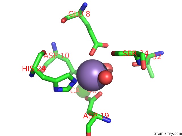
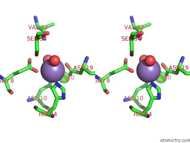
Manganese binding site 1 out of 4 in 5cna
Go back to
Manganese binding site 1 out
of 4 in the Refined Structure of Concanavalin A Complexed with Alpha- Methyl-D-Mannopyranoside at 2.0 Angstroms Resolution and Comparison with the Saccharide-Free Structure
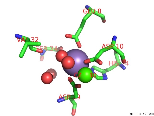
Mono view
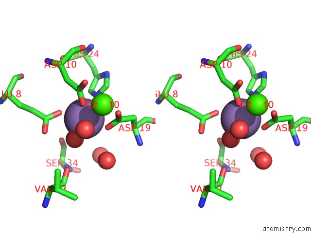
Stereo pair view
Mono view
Stereo pair view
A full contact list of Manganese with other atoms in the Mn binding
site number 1 of Refined Structure of Concanavalin A Complexed with Alpha- Methyl-D-Mannopyranoside at 2.0 Angstroms Resolution and Comparison with the Saccharide-Free Structure within 5.0Å range:
|
Manganese binding site 2 out of 4 in 5cna
Go back to
Manganese binding site 2 out
of 4 in the Refined Structure of Concanavalin A Complexed with Alpha- Methyl-D-Mannopyranoside at 2.0 Angstroms Resolution and Comparison with the Saccharide-Free Structure
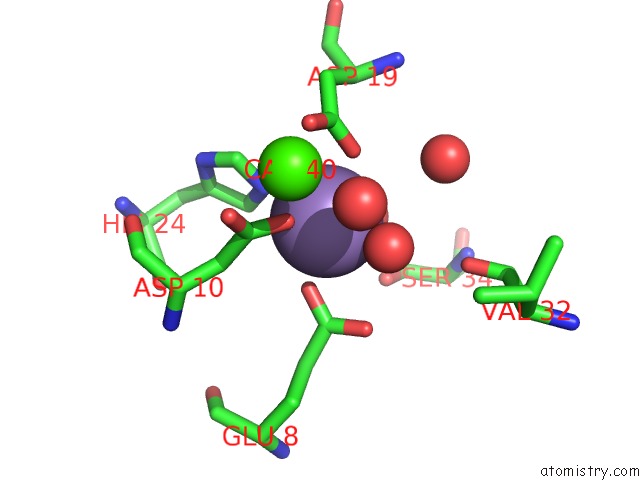
Mono view
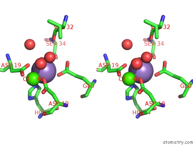
Stereo pair view
Mono view
Stereo pair view
A full contact list of Manganese with other atoms in the Mn binding
site number 2 of Refined Structure of Concanavalin A Complexed with Alpha- Methyl-D-Mannopyranoside at 2.0 Angstroms Resolution and Comparison with the Saccharide-Free Structure within 5.0Å range:
|
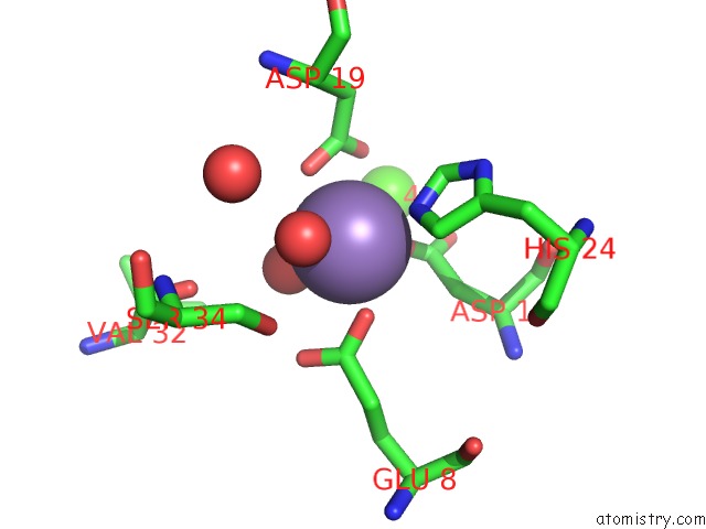
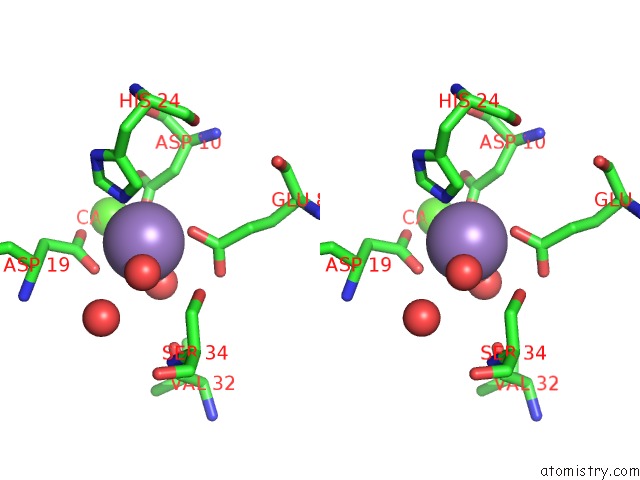
Manganese binding site 3 out of 4 in 5cna
Go back to
Manganese binding site 3 out
of 4 in the Refined Structure of Concanavalin A Complexed with Alpha- Methyl-D-Mannopyranoside at 2.0 Angstroms Resolution and Comparison with the Saccharide-Free Structure
Mono view
Stereo pair view
Mono view
Stereo pair view
A full contact list of Manganese with other atoms in the Mn binding
site number 3 of Refined Structure of Concanavalin A Complexed with Alpha- Methyl-D-Mannopyranoside at 2.0 Angstroms Resolution and Comparison with the Saccharide-Free Structure within 5.0Å range:
|
Manganese binding site 4 out of 4 in 5cna
Go back to
Manganese binding site 4 out
of 4 in the Refined Structure of Concanavalin A Complexed with Alpha- Methyl-D-Mannopyranoside at 2.0 Angstroms Resolution and Comparison with the Saccharide-Free Structure
Mono view
Stereo pair view
Mono view
Stereo pair view
A full contact list of Manganese with other atoms in the Mn binding
site number 4 of Refined Structure of Concanavalin A Complexed with Alpha- Methyl-D-Mannopyranoside at 2.0 Angstroms Resolution and Comparison with the Saccharide-Free Structure within 5.0Å range:
|
Reference:
J.H.Naismith,
C.Emmerich,
J.Habash,
S.J.Harrop,
J.R.Helliwell,
W.N.Hunter,
J.Raftery,
A.J.Kalb,
J.Yariv.
Refined Structure of Concanavalin A Complexed with Methyl Alpha-D-Mannopyranoside at 2.0 A Resolution and Comparison with the Saccharide-Free Structure. Acta Crystallogr.,Sect.D V. 50 847 1994.
ISSN: ISSN 0907-4449
PubMed: 15299352
DOI: 10.1107/S0907444994005287
Page generated: Sat Oct 5 23:47:45 2024
ISSN: ISSN 0907-4449
PubMed: 15299352
DOI: 10.1107/S0907444994005287
Last articles
K in 2VXYK in 2VQW
K in 2VQV
K in 2VQQ
K in 2VQO
K in 2VI5
K in 2VQM
K in 2VQJ
K in 2VPL
K in 2VLH