Manganese »
PDB 5cdn-5dcb »
5cdn »
Manganese in PDB 5cdn: 2.8A Structure of Etoposide with S.Aureus Dna Gyrase and Dna
Enzymatic activity of 2.8A Structure of Etoposide with S.Aureus Dna Gyrase and Dna
All present enzymatic activity of 2.8A Structure of Etoposide with S.Aureus Dna Gyrase and Dna:
5.99.1.3;
5.99.1.3;
Protein crystallography data
The structure of 2.8A Structure of Etoposide with S.Aureus Dna Gyrase and Dna, PDB code: 5cdn
was solved by
B.D.Bax,
V.Srikannathasan,
P.F.Chan,
with X-Ray Crystallography technique. A brief refinement statistics is given in the table below:
| Resolution Low / High (Å) | 39.35 / 2.79 |
| Space group | P 1 21 1 |
| Cell size a, b, c (Å), α, β, γ (°) | 89.782, 170.081, 124.540, 90.00, 102.27, 90.00 |
| R / Rfree (%) | 21.3 / 24.2 |
Other elements in 5cdn:
The structure of 2.8A Structure of Etoposide with S.Aureus Dna Gyrase and Dna also contains other interesting chemical elements:
| Sodium | (Na) | 1 atom |
Manganese Binding Sites:
The binding sites of Manganese atom in the 2.8A Structure of Etoposide with S.Aureus Dna Gyrase and Dna
(pdb code 5cdn). This binding sites where shown within
5.0 Angstroms radius around Manganese atom.
In total 4 binding sites of Manganese where determined in the 2.8A Structure of Etoposide with S.Aureus Dna Gyrase and Dna, PDB code: 5cdn:
Jump to Manganese binding site number: 1; 2; 3; 4;
In total 4 binding sites of Manganese where determined in the 2.8A Structure of Etoposide with S.Aureus Dna Gyrase and Dna, PDB code: 5cdn:
Jump to Manganese binding site number: 1; 2; 3; 4;
Manganese binding site 1 out of 4 in 5cdn
Go back to Manganese Binding Sites List in 5cdn
Manganese binding site 1 out
of 4 in the 2.8A Structure of Etoposide with S.Aureus Dna Gyrase and Dna
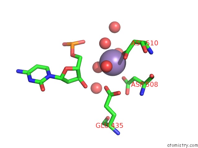
Mono view
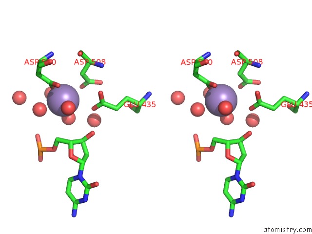
Stereo pair view
Mono view
Stereo pair view
A full contact list of Manganese with other atoms in the Mn binding
site number 1 of 2.8A Structure of Etoposide with S.Aureus Dna Gyrase and Dna within 5.0Å range:
|
Manganese binding site 2 out of 4 in 5cdn
Go back to
Manganese Binding Sites List in 5cdn
Manganese binding site 2 out
of 4 in the 2.8A Structure of Etoposide with S.Aureus Dna Gyrase and Dna
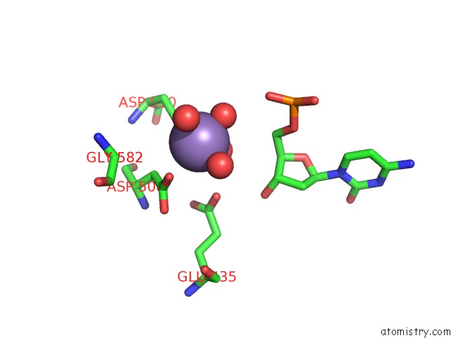
Mono view
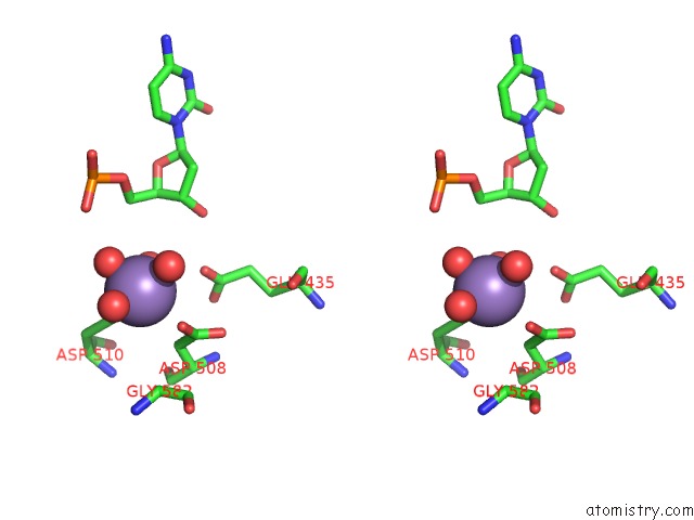
Stereo pair view
Mono view
Stereo pair view
A full contact list of Manganese with other atoms in the Mn binding
site number 2 of 2.8A Structure of Etoposide with S.Aureus Dna Gyrase and Dna within 5.0Å range:
|
Manganese binding site 3 out of 4 in 5cdn
Go back to
Manganese Binding Sites List in 5cdn
Manganese binding site 3 out
of 4 in the 2.8A Structure of Etoposide with S.Aureus Dna Gyrase and Dna
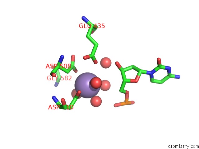
Mono view
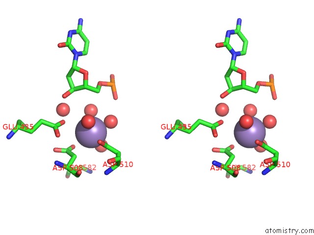
Stereo pair view
Mono view
Stereo pair view
A full contact list of Manganese with other atoms in the Mn binding
site number 3 of 2.8A Structure of Etoposide with S.Aureus Dna Gyrase and Dna within 5.0Å range:
|
Manganese binding site 4 out of 4 in 5cdn
Go back to
Manganese Binding Sites List in 5cdn
Manganese binding site 4 out
of 4 in the 2.8A Structure of Etoposide with S.Aureus Dna Gyrase and Dna
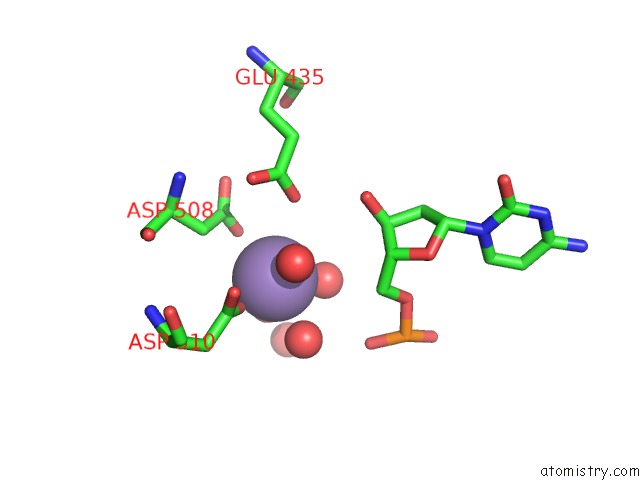
Mono view
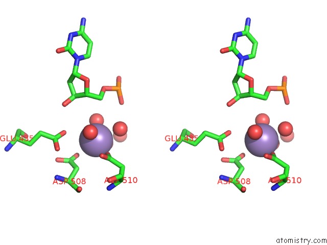
Stereo pair view
Mono view
Stereo pair view
A full contact list of Manganese with other atoms in the Mn binding
site number 4 of 2.8A Structure of Etoposide with S.Aureus Dna Gyrase and Dna within 5.0Å range:
|
Reference:
P.F.Chan,
V.Srikannathasan,
J.Huang,
H.Cui,
A.P.Fosberry,
M.Gu,
M.M.Hann,
M.Hibbs,
P.Homes,
K.Ingraham,
J.Pizzollo,
C.Shen,
A.J.Shillings,
C.E.Spitzfaden,
R.Tanner,
A.J.Theobald,
R.A.Stavenger,
B.D.Bax,
M.N.Gwynn.
Structural Basis of Dna Gyrase Inhibition By Antibacterial Qpt-1, Anticancer Drug Etoposide and Moxifloxacin. Nat Commun V. 6 10048 2015.
ISSN: ESSN 2041-1723
PubMed: 26640131
DOI: 10.1038/NCOMMS10048
Page generated: Sat Oct 5 23:45:20 2024
ISSN: ESSN 2041-1723
PubMed: 26640131
DOI: 10.1038/NCOMMS10048
Last articles
I in 3S2OI in 3RVI
I in 3S2H
I in 3RU1
I in 3S18
I in 3RX6
I in 3RSI
I in 3RG2
I in 3RTN
I in 3RTM
