Manganese »
PDB 4php-4qsf »
4qsf »
Manganese in PDB 4qsf: Crystal Structure of Amidohydrolase PMI1525 (Target Efi-500319) From Proteus Mirabilis HI4320, A Complex with Butyric Acid and Manganese
Protein crystallography data
The structure of Crystal Structure of Amidohydrolase PMI1525 (Target Efi-500319) From Proteus Mirabilis HI4320, A Complex with Butyric Acid and Manganese, PDB code: 4qsf
was solved by
Y.Patskovsky,
R.Toro,
D.F.Xiang,
F.M.Raushel,
J.A.Gerlt,
S.C.Almo,
Enzymefunction Initiative (Efi),
with X-Ray Crystallography technique. A brief refinement statistics is given in the table below:
| Resolution Low / High (Å) | 36.46 / 1.65 |
| Space group | P 32 2 1 |
| Cell size a, b, c (Å), α, β, γ (°) | 101.269, 101.269, 65.493, 90.00, 90.00, 120.00 |
| R / Rfree (%) | 15.5 / 18.1 |
Manganese Binding Sites:
The binding sites of Manganese atom in the Crystal Structure of Amidohydrolase PMI1525 (Target Efi-500319) From Proteus Mirabilis HI4320, A Complex with Butyric Acid and Manganese
(pdb code 4qsf). This binding sites where shown within
5.0 Angstroms radius around Manganese atom.
In total 2 binding sites of Manganese where determined in the Crystal Structure of Amidohydrolase PMI1525 (Target Efi-500319) From Proteus Mirabilis HI4320, A Complex with Butyric Acid and Manganese, PDB code: 4qsf:
Jump to Manganese binding site number: 1; 2;
In total 2 binding sites of Manganese where determined in the Crystal Structure of Amidohydrolase PMI1525 (Target Efi-500319) From Proteus Mirabilis HI4320, A Complex with Butyric Acid and Manganese, PDB code: 4qsf:
Jump to Manganese binding site number: 1; 2;
Manganese binding site 1 out of 2 in 4qsf
Go back to
Manganese binding site 1 out
of 2 in the Crystal Structure of Amidohydrolase PMI1525 (Target Efi-500319) From Proteus Mirabilis HI4320, A Complex with Butyric Acid and Manganese

Mono view
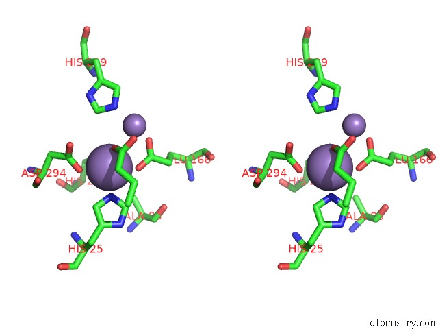
Stereo pair view
Mono view
Stereo pair view
A full contact list of Manganese with other atoms in the Mn binding
site number 1 of Crystal Structure of Amidohydrolase PMI1525 (Target Efi-500319) From Proteus Mirabilis HI4320, A Complex with Butyric Acid and Manganese within 5.0Å range:
|
Manganese binding site 2 out of 2 in 4qsf
Go back to
Manganese binding site 2 out
of 2 in the Crystal Structure of Amidohydrolase PMI1525 (Target Efi-500319) From Proteus Mirabilis HI4320, A Complex with Butyric Acid and Manganese

Mono view

Stereo pair view
Mono view
Stereo pair view
A full contact list of Manganese with other atoms in the Mn binding
site number 2 of Crystal Structure of Amidohydrolase PMI1525 (Target Efi-500319) From Proteus Mirabilis HI4320, A Complex with Butyric Acid and Manganese within 5.0Å range:
|
Reference:
Y.Patskovsky,
R.Toro,
D.F.Xiang,
F.M.Raushel,
S.C.Almo.
Crystal Structure of Amidohydrolase PMI1525 From Proteus Mirabilis HI4320 To Be Published.
Page generated: Sat Oct 5 21:03:06 2024
Last articles
K in 6GEPK in 6G6R
K in 6G2M
K in 6FSH
K in 6FQ2
K in 6FQD
K in 6FI0
K in 6FHK
K in 6F3O
K in 6FCD