Manganese »
PDB 3x30-4b5h »
3zy2 »
Manganese in PDB 3zy2: Crystal Structure of POFUT1 in Complex with Gdp (High Resolution Dataset)
Enzymatic activity of Crystal Structure of POFUT1 in Complex with Gdp (High Resolution Dataset)
All present enzymatic activity of Crystal Structure of POFUT1 in Complex with Gdp (High Resolution Dataset):
2.4.1.221;
2.4.1.221;
Protein crystallography data
The structure of Crystal Structure of POFUT1 in Complex with Gdp (High Resolution Dataset), PDB code: 3zy2
was solved by
E.Lira-Navarrete,
J.Valero-Gonzalez,
R.Villanueva,
M.Martinez-Julvez,
T.Tejero,
P.Merino,
S.Panjikar,
R.Hurtado-Guerrero,
with X-Ray Crystallography technique. A brief refinement statistics is given in the table below:
| Resolution Low / High (Å) | 70.63 / 1.54 |
| Space group | C 1 2 1 |
| Cell size a, b, c (Å), α, β, γ (°) | 145.018, 38.247, 68.196, 90.00, 103.07, 90.00 |
| R / Rfree (%) | 20.507 / 23.725 |
Manganese Binding Sites:
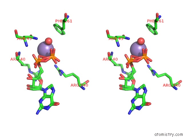
The binding sites of Manganese atom in the Crystal Structure of POFUT1 in Complex with Gdp (High Resolution Dataset)
(pdb code 3zy2). This binding sites where shown within
5.0 Angstroms radius around Manganese atom.
In total only one binding site of Manganese was determined in the Crystal Structure of POFUT1 in Complex with Gdp (High Resolution Dataset), PDB code: 3zy2:
In total only one binding site of Manganese was determined in the Crystal Structure of POFUT1 in Complex with Gdp (High Resolution Dataset), PDB code: 3zy2:
Manganese binding site 1 out of 1 in 3zy2
Go back to
Manganese binding site 1 out
of 1 in the Crystal Structure of POFUT1 in Complex with Gdp (High Resolution Dataset)

Mono view
Stereo pair view
Mono view
Stereo pair view
A full contact list of Manganese with other atoms in the Mn binding
site number 1 of Crystal Structure of POFUT1 in Complex with Gdp (High Resolution Dataset) within 5.0Å range:
|
Reference:
E.Lira-Navarrete,
J.Valero-Gonzalez,
R.Villanueva,
M.Martinez-Julvez,
T.Tejero,
P.Merino,
S.Panjikar,
R.Hurtado-Guerrero.
Structural Insights Into the Mechanism of Protein O-Fucosylation. Plos One V. 6 25365 2011.
ISSN: ISSN 1932-6203
PubMed: 21966509
DOI: 10.1371/JOURNAL.PONE.0025365
Page generated: Sat Oct 5 18:42:52 2024
ISSN: ISSN 1932-6203
PubMed: 21966509
DOI: 10.1371/JOURNAL.PONE.0025365
Last articles
Mg in 3G4TMg in 3G4L
Mg in 3G4K
Mg in 3G4I
Mg in 3G4G
Mg in 3G4F
Mg in 3G45
Mg in 3G3Y
Mg in 3G2X
Mg in 3G3P