Manganese »
PDB 2hvh-2jcj »
2j0b »
Manganese in PDB 2j0b: Structure of the Catalytic Domain of Mouse Manic Fringe in Complex with Udp and Manganese
Enzymatic activity of Structure of the Catalytic Domain of Mouse Manic Fringe in Complex with Udp and Manganese
All present enzymatic activity of Structure of the Catalytic Domain of Mouse Manic Fringe in Complex with Udp and Manganese:
2.4.1.222;
2.4.1.222;
Protein crystallography data
The structure of Structure of the Catalytic Domain of Mouse Manic Fringe in Complex with Udp and Manganese, PDB code: 2j0b
was solved by
M.Jinek,
Y.-W.Chen,
H.Clausen,
S.M.Cohen,
E.Conti,
with X-Ray Crystallography technique. A brief refinement statistics is given in the table below:
| Resolution Low / High (Å) | 80.85 / 2.10 |
| Space group | P 21 21 2 |
| Cell size a, b, c (Å), α, β, γ (°) | 161.760, 40.970, 38.370, 90.00, 90.00, 90.00 |
| R / Rfree (%) | 17.7 / 22.1 |
Other elements in 2j0b:
The structure of Structure of the Catalytic Domain of Mouse Manic Fringe in Complex with Udp and Manganese also contains other interesting chemical elements:
| Potassium | (K) | 1 atom |
Manganese Binding Sites:
The binding sites of Manganese atom in the Structure of the Catalytic Domain of Mouse Manic Fringe in Complex with Udp and Manganese
(pdb code 2j0b). This binding sites where shown within
5.0 Angstroms radius around Manganese atom.
In total only one binding site of Manganese was determined in the Structure of the Catalytic Domain of Mouse Manic Fringe in Complex with Udp and Manganese, PDB code: 2j0b:
In total only one binding site of Manganese was determined in the Structure of the Catalytic Domain of Mouse Manic Fringe in Complex with Udp and Manganese, PDB code: 2j0b:
Manganese binding site 1 out of 1 in 2j0b
Go back to
Manganese binding site 1 out
of 1 in the Structure of the Catalytic Domain of Mouse Manic Fringe in Complex with Udp and Manganese
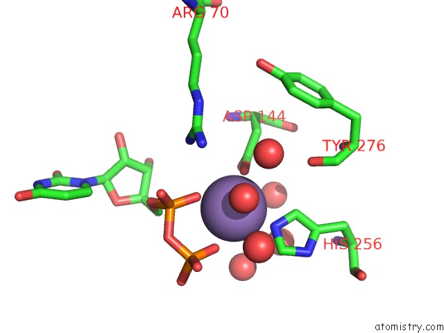
Mono view
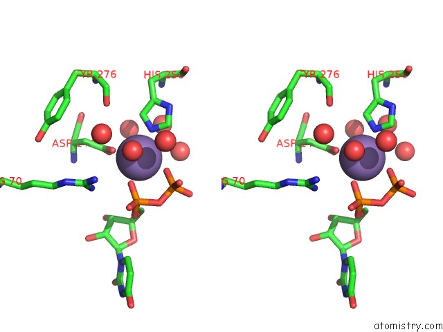
Stereo pair view
Mono view
Stereo pair view
A full contact list of Manganese with other atoms in the Mn binding
site number 1 of Structure of the Catalytic Domain of Mouse Manic Fringe in Complex with Udp and Manganese within 5.0Å range:
|
Reference:
M.Jinek,
Y.-W.Chen,
H.Clausen,
S.M.Cohen,
E.Conti.
Structural Insights Into the Notch-Modifying Glycosyltransferase Fringe Nat.Struct.Mol.Biol. V. 13 945 2006.
ISSN: ISSN 1545-9993
PubMed: 16964258
DOI: 10.1038/NSMB1144
Page generated: Sat Aug 16 10:29:08 2025
ISSN: ISSN 1545-9993
PubMed: 16964258
DOI: 10.1038/NSMB1144
Last articles
Na in 1KCCNa in 1KC6
Na in 1KA0
Na in 1K8Y
Na in 1K8Z
Na in 1K8X
Na in 1K7X
Na in 1K7E
Na in 1K8P
Na in 1K73