Manganese »
PDB 2hvh-2jcj »
2irx »
Manganese in PDB 2irx: Crystal Structure of the Polymerase Domain From Mycobacterium Tuberculosis Ligase D with Gtp and Manganese.
Protein crystallography data
The structure of Crystal Structure of the Polymerase Domain From Mycobacterium Tuberculosis Ligase D with Gtp and Manganese., PDB code: 2irx
was solved by
N.C.Brissett,
R.S.Pitcher,
A.J.Doherty,
with X-Ray Crystallography technique. A brief refinement statistics is given in the table below:
| Resolution Low / High (Å) | 34.79 / 1.80 |
| Space group | P 21 21 21 |
| Cell size a, b, c (Å), α, β, γ (°) | 42.557, 75.532, 89.518, 90.00, 90.00, 90.00 |
| R / Rfree (%) | 16.4 / 20.9 |
Manganese Binding Sites:
The binding sites of Manganese atom in the Crystal Structure of the Polymerase Domain From Mycobacterium Tuberculosis Ligase D with Gtp and Manganese.
(pdb code 2irx). This binding sites where shown within
5.0 Angstroms radius around Manganese atom.
In total only one binding site of Manganese was determined in the Crystal Structure of the Polymerase Domain From Mycobacterium Tuberculosis Ligase D with Gtp and Manganese., PDB code: 2irx:
In total only one binding site of Manganese was determined in the Crystal Structure of the Polymerase Domain From Mycobacterium Tuberculosis Ligase D with Gtp and Manganese., PDB code: 2irx:
Manganese binding site 1 out of 1 in 2irx
Go back to
Manganese binding site 1 out
of 1 in the Crystal Structure of the Polymerase Domain From Mycobacterium Tuberculosis Ligase D with Gtp and Manganese.
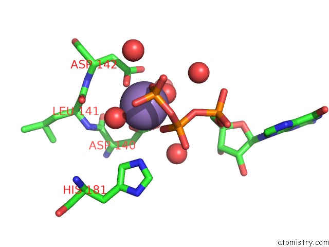
Mono view
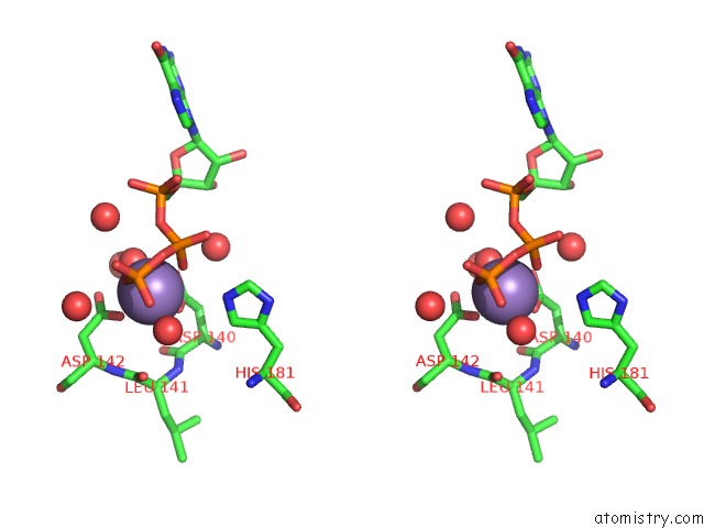
Stereo pair view
Mono view
Stereo pair view
A full contact list of Manganese with other atoms in the Mn binding
site number 1 of Crystal Structure of the Polymerase Domain From Mycobacterium Tuberculosis Ligase D with Gtp and Manganese. within 5.0Å range:
|
Reference:
R.S.Pitcher,
N.C.Brissett,
A.J.Picher,
P.Andrade,
R.Juarez,
D.Thompson,
G.C.Fox,
L.Blanco,
A.J.Doherty.
Structure and Function of A Mycobacterial Nhej Dna Repair Polymerase. J.Mol.Biol. V. 366 391 2007.
ISSN: ISSN 0022-2836
PubMed: 17174332
DOI: 10.1016/J.JMB.2006.10.046
Page generated: Sat Oct 5 14:29:57 2024
ISSN: ISSN 0022-2836
PubMed: 17174332
DOI: 10.1016/J.JMB.2006.10.046
Last articles
Mg in 3IJQMg in 3IJL
Mg in 3IJI
Mg in 3IJH
Mg in 3IIV
Mg in 3IIU
Mg in 3IIL
Mg in 3IIS
Mg in 3IHK
Mg in 3II9