Manganese »
PDB 5ekw-5fxv »
5fh4 »
Manganese in PDB 5fh4: The Structure of Rat Cytosolic Pepck Variant E89D in Complex with Beta-Sulfopyruvate and Gtp
Enzymatic activity of The Structure of Rat Cytosolic Pepck Variant E89D in Complex with Beta-Sulfopyruvate and Gtp
All present enzymatic activity of The Structure of Rat Cytosolic Pepck Variant E89D in Complex with Beta-Sulfopyruvate and Gtp:
4.1.1.32;
4.1.1.32;
Protein crystallography data
The structure of The Structure of Rat Cytosolic Pepck Variant E89D in Complex with Beta-Sulfopyruvate and Gtp, PDB code: 5fh4
was solved by
T.A.Johnson,
T.Holyoak,
with X-Ray Crystallography technique. A brief refinement statistics is given in the table below:
| Resolution Low / High (Å) | 34.34 / 1.49 |
| Space group | P 1 21 1 |
| Cell size a, b, c (Å), α, β, γ (°) | 44.590, 118.809, 60.508, 90.00, 109.30, 90.00 |
| R / Rfree (%) | 20.4 / 22.9 |
Other elements in 5fh4:
The structure of The Structure of Rat Cytosolic Pepck Variant E89D in Complex with Beta-Sulfopyruvate and Gtp also contains other interesting chemical elements:
| Sodium | (Na) | 1 atom |
Manganese Binding Sites:
The binding sites of Manganese atom in the The Structure of Rat Cytosolic Pepck Variant E89D in Complex with Beta-Sulfopyruvate and Gtp
(pdb code 5fh4). This binding sites where shown within
5.0 Angstroms radius around Manganese atom.
In total 3 binding sites of Manganese where determined in the The Structure of Rat Cytosolic Pepck Variant E89D in Complex with Beta-Sulfopyruvate and Gtp, PDB code: 5fh4:
Jump to Manganese binding site number: 1; 2; 3;
In total 3 binding sites of Manganese where determined in the The Structure of Rat Cytosolic Pepck Variant E89D in Complex with Beta-Sulfopyruvate and Gtp, PDB code: 5fh4:
Jump to Manganese binding site number: 1; 2; 3;
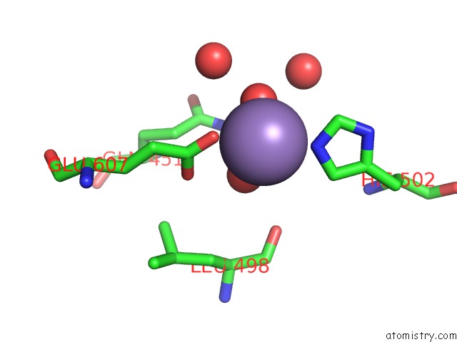
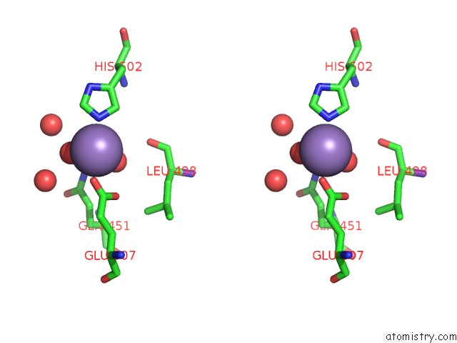
Manganese binding site 1 out of 3 in 5fh4
Go back to
Manganese binding site 1 out
of 3 in the The Structure of Rat Cytosolic Pepck Variant E89D in Complex with Beta-Sulfopyruvate and Gtp
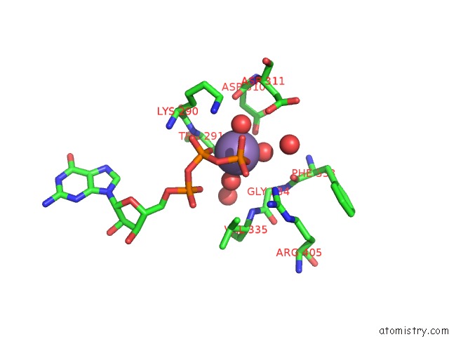
Mono view
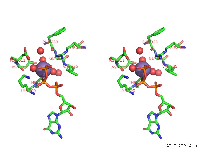
Stereo pair view
Mono view
Stereo pair view
A full contact list of Manganese with other atoms in the Mn binding
site number 1 of The Structure of Rat Cytosolic Pepck Variant E89D in Complex with Beta-Sulfopyruvate and Gtp within 5.0Å range:
|
Manganese binding site 2 out of 3 in 5fh4
Go back to
Manganese binding site 2 out
of 3 in the The Structure of Rat Cytosolic Pepck Variant E89D in Complex with Beta-Sulfopyruvate and Gtp
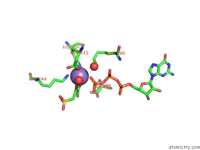
Mono view
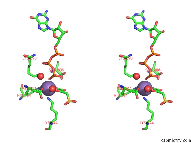
Stereo pair view
Mono view
Stereo pair view
A full contact list of Manganese with other atoms in the Mn binding
site number 2 of The Structure of Rat Cytosolic Pepck Variant E89D in Complex with Beta-Sulfopyruvate and Gtp within 5.0Å range:
|
Manganese binding site 3 out of 3 in 5fh4
Go back to
Manganese binding site 3 out
of 3 in the The Structure of Rat Cytosolic Pepck Variant E89D in Complex with Beta-Sulfopyruvate and Gtp
Mono view
Stereo pair view
Mono view
Stereo pair view
A full contact list of Manganese with other atoms in the Mn binding
site number 3 of The Structure of Rat Cytosolic Pepck Variant E89D in Complex with Beta-Sulfopyruvate and Gtp within 5.0Å range:
|
Reference:
T.A.Johnson,
M.J.Mcleod,
T.Holyoak.
Utilization of Substrate Intrinsic Binding Energy For Conformational Change and Catalytic Function in Phosphoenolpyruvate Carboxykinase. Biochemistry V. 55 575 2016.
ISSN: ISSN 0006-2960
PubMed: 26709450
DOI: 10.1021/ACS.BIOCHEM.5B01215
Page generated: Sat Aug 16 17:24:13 2025
ISSN: ISSN 0006-2960
PubMed: 26709450
DOI: 10.1021/ACS.BIOCHEM.5B01215
Last articles
Mn in 9XIMMn in 9XIA
Mn in 9NJO
Mn in 9QB8
Mn in 9MR6
Mn in 9M2Q
Mn in 9NCZ
Mn in 9NB9
Mn in 9M2R
Mn in 9M2P