Manganese »
PDB 5dcd-5ekd »
5eji »
Manganese in PDB 5eji: Crystal Structure of Nad Kinase W78F Mutant From Listeria Monocytogenes in Complex with Nadp/Mn++/Ppi
Enzymatic activity of Crystal Structure of Nad Kinase W78F Mutant From Listeria Monocytogenes in Complex with Nadp/Mn++/Ppi
All present enzymatic activity of Crystal Structure of Nad Kinase W78F Mutant From Listeria Monocytogenes in Complex with Nadp/Mn++/Ppi:
2.7.1.23;
2.7.1.23;
Protein crystallography data
The structure of Crystal Structure of Nad Kinase W78F Mutant From Listeria Monocytogenes in Complex with Nadp/Mn++/Ppi, PDB code: 5eji
was solved by
G.Poncet-Montange,
L.Assairi,
M.Gelin,
S.Pochet,
G.Labesse,
with X-Ray Crystallography technique. A brief refinement statistics is given in the table below:
| Resolution Low / High (Å) | 63.08 / 2.29 |
| Space group | I 2 2 2 |
| Cell size a, b, c (Å), α, β, γ (°) | 63.466, 74.389, 119.012, 90.00, 90.00, 90.00 |
| R / Rfree (%) | 17.7 / 23.7 |
Manganese Binding Sites:
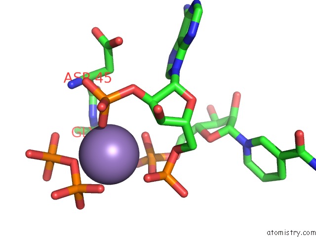
The binding sites of Manganese atom in the Crystal Structure of Nad Kinase W78F Mutant From Listeria Monocytogenes in Complex with Nadp/Mn++/Ppi
(pdb code 5eji). This binding sites where shown within
5.0 Angstroms radius around Manganese atom.
In total only one binding site of Manganese was determined in the Crystal Structure of Nad Kinase W78F Mutant From Listeria Monocytogenes in Complex with Nadp/Mn++/Ppi, PDB code: 5eji:
In total only one binding site of Manganese was determined in the Crystal Structure of Nad Kinase W78F Mutant From Listeria Monocytogenes in Complex with Nadp/Mn++/Ppi, PDB code: 5eji:
Manganese binding site 1 out of 1 in 5eji
Go back to
Manganese binding site 1 out
of 1 in the Crystal Structure of Nad Kinase W78F Mutant From Listeria Monocytogenes in Complex with Nadp/Mn++/Ppi
Mono view
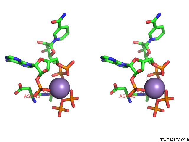
Stereo pair view
Mono view
Stereo pair view
A full contact list of Manganese with other atoms in the Mn binding
site number 1 of Crystal Structure of Nad Kinase W78F Mutant From Listeria Monocytogenes in Complex with Nadp/Mn++/Ppi within 5.0Å range:
|
Reference:
G.Poncet-Montange,
L.Assairi,
M.Gelin,
S.Pochet,
G.Labesse.
Crystal Structure of Nad Kinase W78F Mutant From Listeria Monocytogenes in Complex with Nadp/Mn++/Ppi To Be Published.
Page generated: Sun Oct 6 00:04:42 2024
Last articles
F in 7LCRF in 7LCM
F in 7LCO
F in 7LCK
F in 7LCJ
F in 7LCI
F in 7L9Y
F in 7LCD
F in 7LAY
F in 7LAJ