Manganese »
PDB 2a7a-2axt »
2a8p »
Manganese in PDB 2a8p: 2.7 Angstrom Crystal Structure of the Complex Between the Nuclear Snorna Decapping Nudix Hydrolase X29 and Manganese
Protein crystallography data
The structure of 2.7 Angstrom Crystal Structure of the Complex Between the Nuclear Snorna Decapping Nudix Hydrolase X29 and Manganese, PDB code: 2a8p
was solved by
J.N.Scarsdale,
B.A.Peculis,
H.T.Wright,
with X-Ray Crystallography technique. A brief refinement statistics is given in the table below:
| Resolution Low / High (Å) | 27.60 / 2.70 |
| Space group | P 21 21 21 |
| Cell size a, b, c (Å), α, β, γ (°) | 50.239, 82.045, 112.009, 90.00, 90.00, 90.00 |
| R / Rfree (%) | 19.8 / 26.2 |
Manganese Binding Sites:
The binding sites of Manganese atom in the 2.7 Angstrom Crystal Structure of the Complex Between the Nuclear Snorna Decapping Nudix Hydrolase X29 and Manganese
(pdb code 2a8p). This binding sites where shown within
5.0 Angstroms radius around Manganese atom.
In total 5 binding sites of Manganese where determined in the 2.7 Angstrom Crystal Structure of the Complex Between the Nuclear Snorna Decapping Nudix Hydrolase X29 and Manganese, PDB code: 2a8p:
Jump to Manganese binding site number: 1; 2; 3; 4; 5;
In total 5 binding sites of Manganese where determined in the 2.7 Angstrom Crystal Structure of the Complex Between the Nuclear Snorna Decapping Nudix Hydrolase X29 and Manganese, PDB code: 2a8p:
Jump to Manganese binding site number: 1; 2; 3; 4; 5;
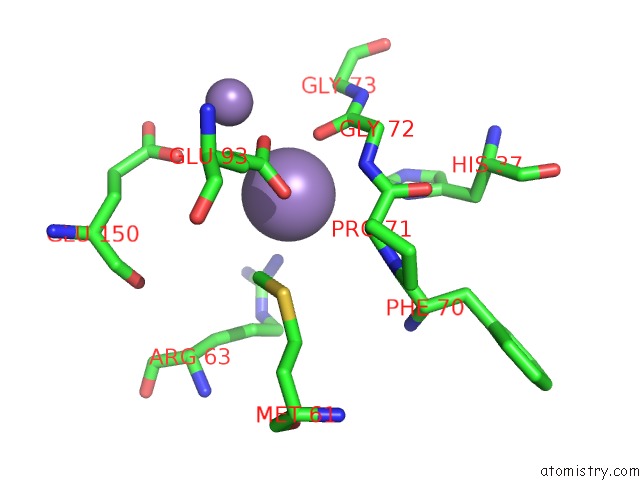
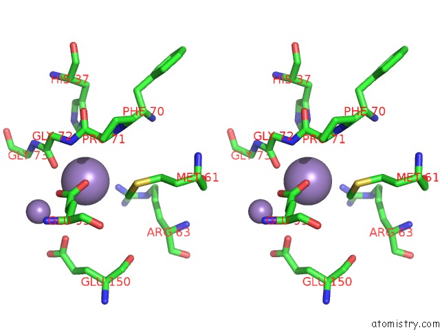
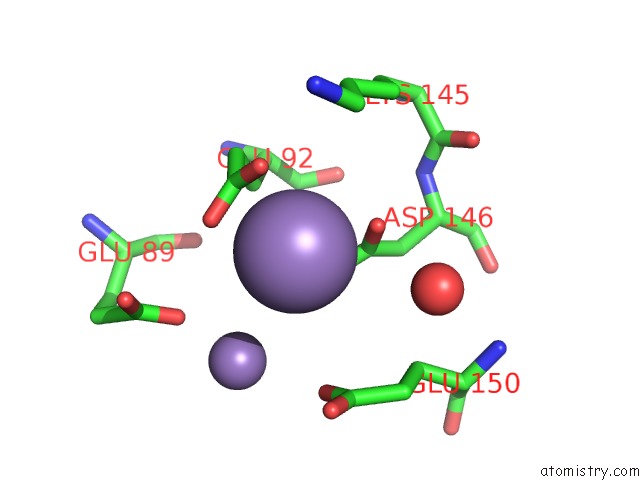
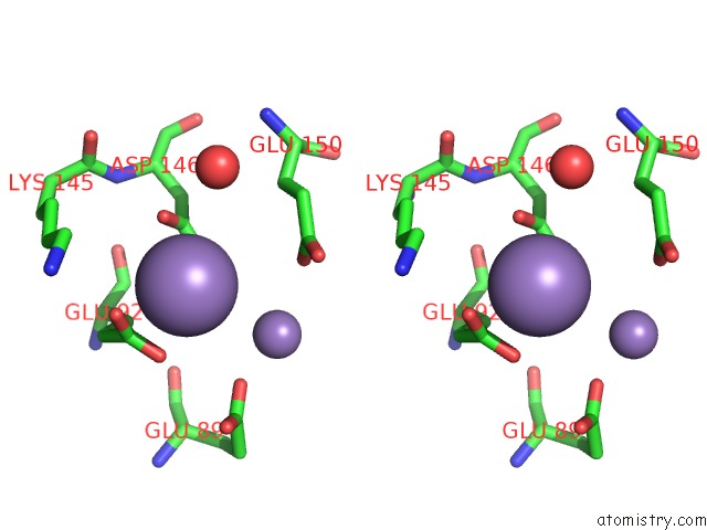
Manganese binding site 1 out of 5 in 2a8p
Go back to
Manganese binding site 1 out
of 5 in the 2.7 Angstrom Crystal Structure of the Complex Between the Nuclear Snorna Decapping Nudix Hydrolase X29 and Manganese
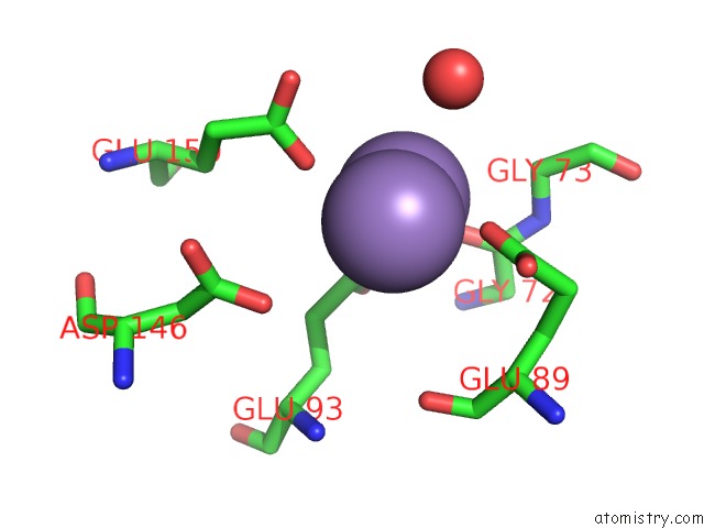
Mono view
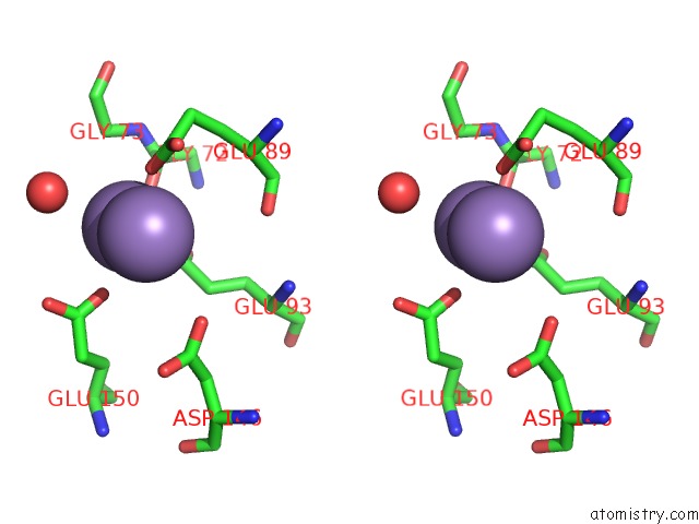
Stereo pair view
Mono view
Stereo pair view
A full contact list of Manganese with other atoms in the Mn binding
site number 1 of 2.7 Angstrom Crystal Structure of the Complex Between the Nuclear Snorna Decapping Nudix Hydrolase X29 and Manganese within 5.0Å range:
|
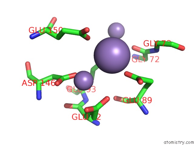
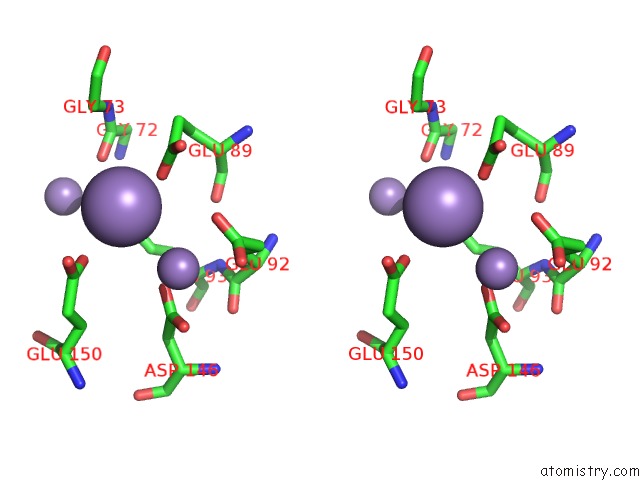
Manganese binding site 2 out of 5 in 2a8p
Go back to
Manganese binding site 2 out
of 5 in the 2.7 Angstrom Crystal Structure of the Complex Between the Nuclear Snorna Decapping Nudix Hydrolase X29 and Manganese
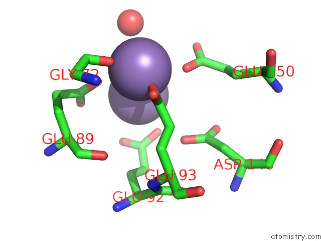
Mono view
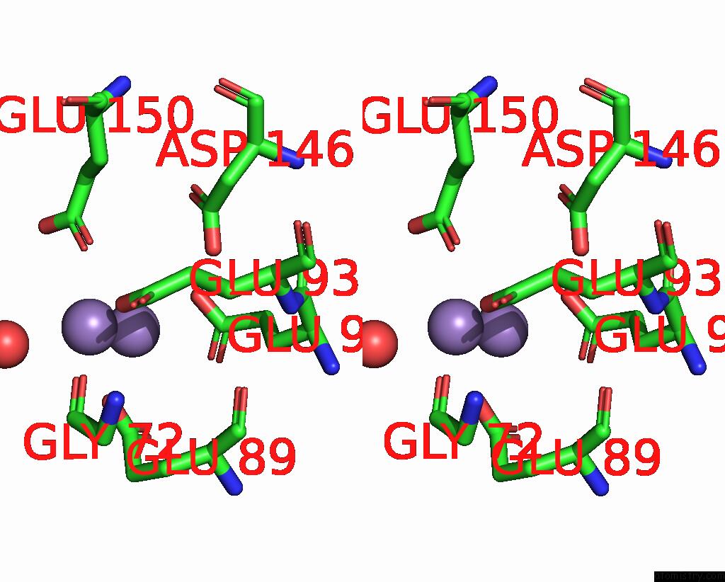
Stereo pair view
Mono view
Stereo pair view
A full contact list of Manganese with other atoms in the Mn binding
site number 2 of 2.7 Angstrom Crystal Structure of the Complex Between the Nuclear Snorna Decapping Nudix Hydrolase X29 and Manganese within 5.0Å range:
|
Manganese binding site 3 out of 5 in 2a8p
Go back to
Manganese binding site 3 out
of 5 in the 2.7 Angstrom Crystal Structure of the Complex Between the Nuclear Snorna Decapping Nudix Hydrolase X29 and Manganese
Mono view
Stereo pair view
Mono view
Stereo pair view
A full contact list of Manganese with other atoms in the Mn binding
site number 3 of 2.7 Angstrom Crystal Structure of the Complex Between the Nuclear Snorna Decapping Nudix Hydrolase X29 and Manganese within 5.0Å range:
|
Manganese binding site 4 out of 5 in 2a8p
Go back to
Manganese binding site 4 out
of 5 in the 2.7 Angstrom Crystal Structure of the Complex Between the Nuclear Snorna Decapping Nudix Hydrolase X29 and Manganese
Mono view
Stereo pair view
Mono view
Stereo pair view
A full contact list of Manganese with other atoms in the Mn binding
site number 4 of 2.7 Angstrom Crystal Structure of the Complex Between the Nuclear Snorna Decapping Nudix Hydrolase X29 and Manganese within 5.0Å range:
|
Manganese binding site 5 out of 5 in 2a8p
Go back to
Manganese binding site 5 out
of 5 in the 2.7 Angstrom Crystal Structure of the Complex Between the Nuclear Snorna Decapping Nudix Hydrolase X29 and Manganese
Mono view
Stereo pair view
Mono view
Stereo pair view
A full contact list of Manganese with other atoms in the Mn binding
site number 5 of 2.7 Angstrom Crystal Structure of the Complex Between the Nuclear Snorna Decapping Nudix Hydrolase X29 and Manganese within 5.0Å range:
|
Reference:
J.N.Scarsdale,
B.A.Peculis,
H.T.Wright.
Crystal Structures of U8 Snorna Decapping Nudix Hydrolase, X29, and Its Metal and Cap Complexes Structure V. 14 331 2006.
ISSN: ISSN 0969-2126
PubMed: 16472752
DOI: 10.1016/J.STR.2005.11.010
Page generated: Sat Aug 16 09:40:43 2025
ISSN: ISSN 0969-2126
PubMed: 16472752
DOI: 10.1016/J.STR.2005.11.010
Last articles
Mn in 8JKKMn in 8JNK
Mn in 8JNR
Mn in 8IRI
Mn in 8JGP
Mn in 8JBM
Mn in 8JDZ
Mn in 8JDY
Mn in 8JDX
Mn in 8JDV