Manganese »
PDB 8wq7-9c4d »
8y2l »
Manganese in PDB 8y2l: The Crystal Structure of Glucosyltransferase Tcdb From Clostridioides Difficile
Protein crystallography data
The structure of The Crystal Structure of Glucosyltransferase Tcdb From Clostridioides Difficile, PDB code: 8y2l
was solved by
S.Fan,
X.Wei,
R.Lv,
C.Wang,
M.Tang,
Y.Jin,
Z.Yang,
with X-Ray Crystallography technique. A brief refinement statistics is given in the table below:
| Resolution Low / High (Å) | 49.15 / 3.95 |
| Space group | C 2 2 21 |
| Cell size a, b, c (Å), α, β, γ (°) | 155.166, 268.755, 194.862, 90, 90, 90 |
| R / Rfree (%) | 24.4 / 30 |
Manganese Binding Sites:
The binding sites of Manganese atom in the The Crystal Structure of Glucosyltransferase Tcdb From Clostridioides Difficile
(pdb code 8y2l). This binding sites where shown within
5.0 Angstroms radius around Manganese atom.
In total 3 binding sites of Manganese where determined in the The Crystal Structure of Glucosyltransferase Tcdb From Clostridioides Difficile, PDB code: 8y2l:
Jump to Manganese binding site number: 1; 2; 3;
In total 3 binding sites of Manganese where determined in the The Crystal Structure of Glucosyltransferase Tcdb From Clostridioides Difficile, PDB code: 8y2l:
Jump to Manganese binding site number: 1; 2; 3;
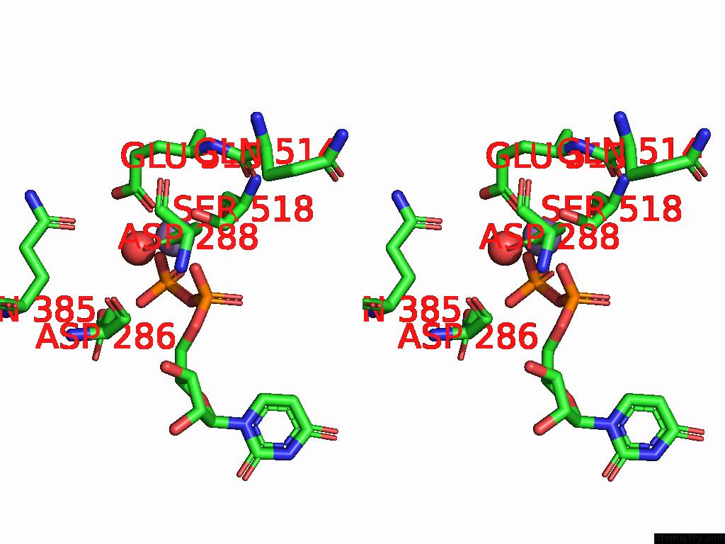
Manganese binding site 1 out of 3 in 8y2l
Go back to
Manganese binding site 1 out
of 3 in the The Crystal Structure of Glucosyltransferase Tcdb From Clostridioides Difficile
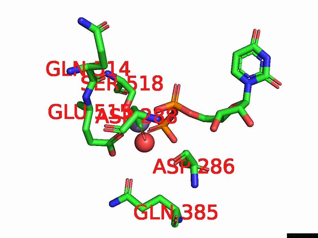
Mono view

Stereo pair view
Mono view
Stereo pair view
A full contact list of Manganese with other atoms in the Mn binding
site number 1 of The Crystal Structure of Glucosyltransferase Tcdb From Clostridioides Difficile within 5.0Å range:
|
Manganese binding site 2 out of 3 in 8y2l
Go back to
Manganese binding site 2 out
of 3 in the The Crystal Structure of Glucosyltransferase Tcdb From Clostridioides Difficile
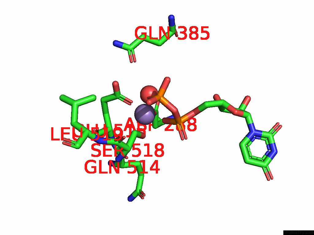
Mono view
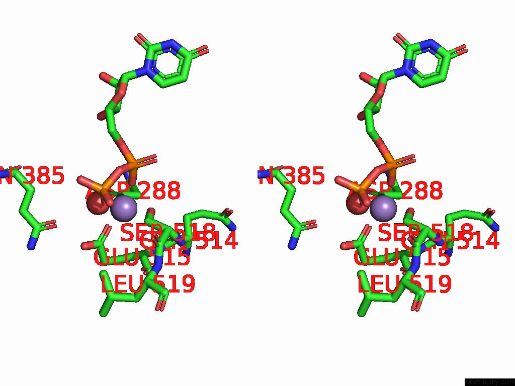
Stereo pair view
Mono view
Stereo pair view
A full contact list of Manganese with other atoms in the Mn binding
site number 2 of The Crystal Structure of Glucosyltransferase Tcdb From Clostridioides Difficile within 5.0Å range:
|
Manganese binding site 3 out of 3 in 8y2l
Go back to
Manganese binding site 3 out
of 3 in the The Crystal Structure of Glucosyltransferase Tcdb From Clostridioides Difficile
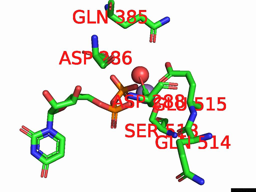
Mono view
Stereo pair view
Mono view
Stereo pair view
A full contact list of Manganese with other atoms in the Mn binding
site number 3 of The Crystal Structure of Glucosyltransferase Tcdb From Clostridioides Difficile within 5.0Å range:
|
Reference:
S.Fan,
X.Wei,
R.Lv,
C.Wang,
M.Tang,
Y.Jin,
Z.Yang.
The Crystal Structure of Glucosyltransferase Tcdb From Clostridioides Difficile To Be Published.
Page generated: Sun Feb 9 08:27:46 2025
Last articles
Mg in 3ZOUMg in 3ZKB
Mg in 3ZOZ
Mg in 3ZOA
Mg in 3ZNU
Mg in 3ZO9
Mg in 3ZM7
Mg in 3ZMC
Mg in 3ZN8
Mg in 3ZKD