Manganese »
PDB 8wq7-9c4d »
8y17 »
Manganese in PDB 8y17: Crystal Structure of A Mutant Staphylococcus Equorum Manganese Superoxide Dismutase T89C and N187C
Enzymatic activity of Crystal Structure of A Mutant Staphylococcus Equorum Manganese Superoxide Dismutase T89C and N187C
All present enzymatic activity of Crystal Structure of A Mutant Staphylococcus Equorum Manganese Superoxide Dismutase T89C and N187C:
1.15.1.1;
1.15.1.1;
Protein crystallography data
The structure of Crystal Structure of A Mutant Staphylococcus Equorum Manganese Superoxide Dismutase T89C and N187C, PDB code: 8y17
was solved by
N.A.Putri,
M.D.Fadillah,
H.Yoshida,
D.S.Retnoningrum,
A.A.Artarini,
R.A.Utami,
W.T.Ismaya,
with X-Ray Crystallography technique. A brief refinement statistics is given in the table below:
| Resolution Low / High (Å) | 47.81 / 2.80 |
| Space group | C 1 2 1 |
| Cell size a, b, c (Å), α, β, γ (°) | 156.92, 135.82, 76.38, 90, 102.74, 90 |
| R / Rfree (%) | 23.4 / 27.6 |
Manganese Binding Sites:
The binding sites of Manganese atom in the Crystal Structure of A Mutant Staphylococcus Equorum Manganese Superoxide Dismutase T89C and N187C
(pdb code 8y17). This binding sites where shown within
5.0 Angstroms radius around Manganese atom.
In total 4 binding sites of Manganese where determined in the Crystal Structure of A Mutant Staphylococcus Equorum Manganese Superoxide Dismutase T89C and N187C, PDB code: 8y17:
Jump to Manganese binding site number: 1; 2; 3; 4;
In total 4 binding sites of Manganese where determined in the Crystal Structure of A Mutant Staphylococcus Equorum Manganese Superoxide Dismutase T89C and N187C, PDB code: 8y17:
Jump to Manganese binding site number: 1; 2; 3; 4;
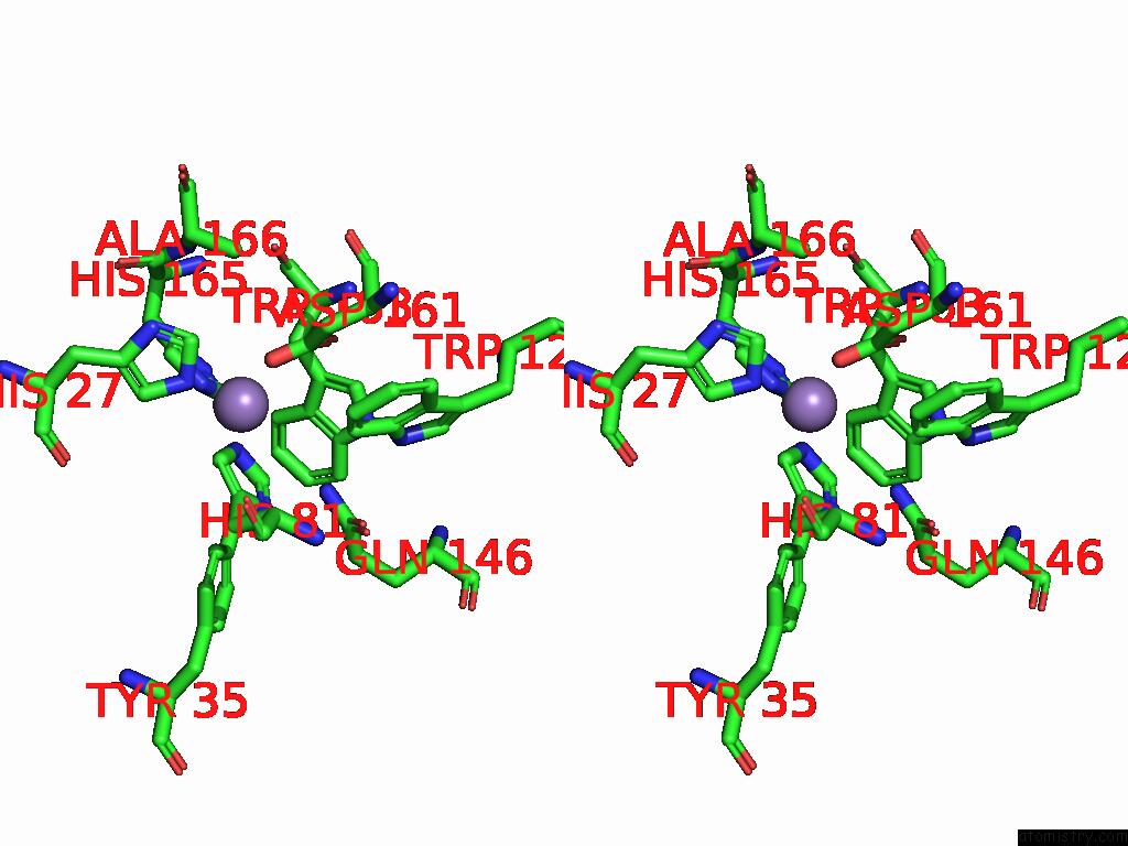
Manganese binding site 1 out of 4 in 8y17
Go back to
Manganese binding site 1 out
of 4 in the Crystal Structure of A Mutant Staphylococcus Equorum Manganese Superoxide Dismutase T89C and N187C
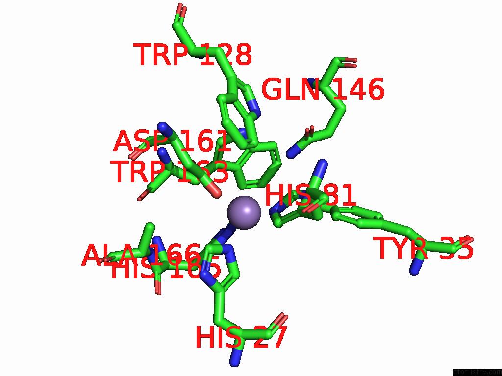
Mono view
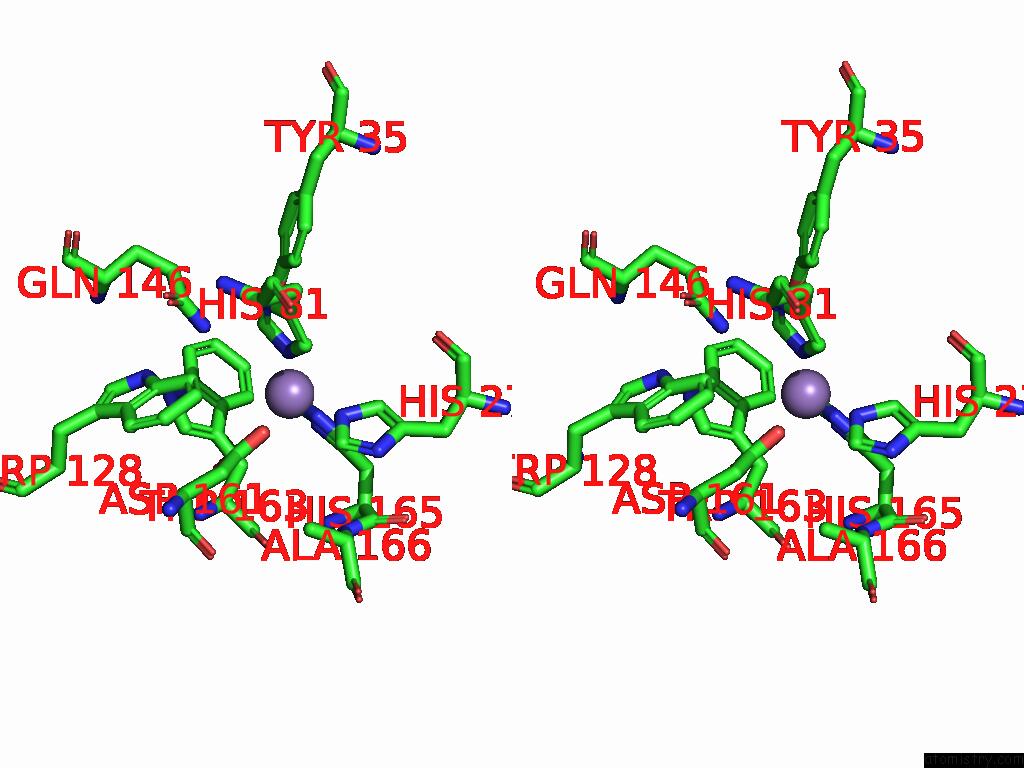
Stereo pair view
Mono view
Stereo pair view
A full contact list of Manganese with other atoms in the Mn binding
site number 1 of Crystal Structure of A Mutant Staphylococcus Equorum Manganese Superoxide Dismutase T89C and N187C within 5.0Å range:
|
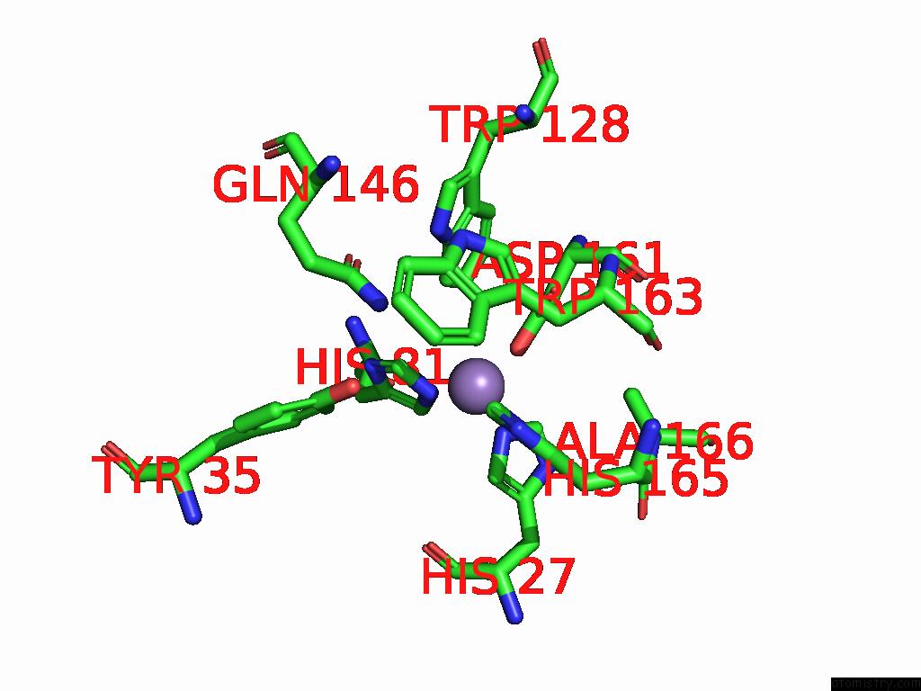
Manganese binding site 2 out of 4 in 8y17
Go back to
Manganese binding site 2 out
of 4 in the Crystal Structure of A Mutant Staphylococcus Equorum Manganese Superoxide Dismutase T89C and N187C
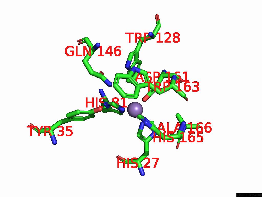
Mono view
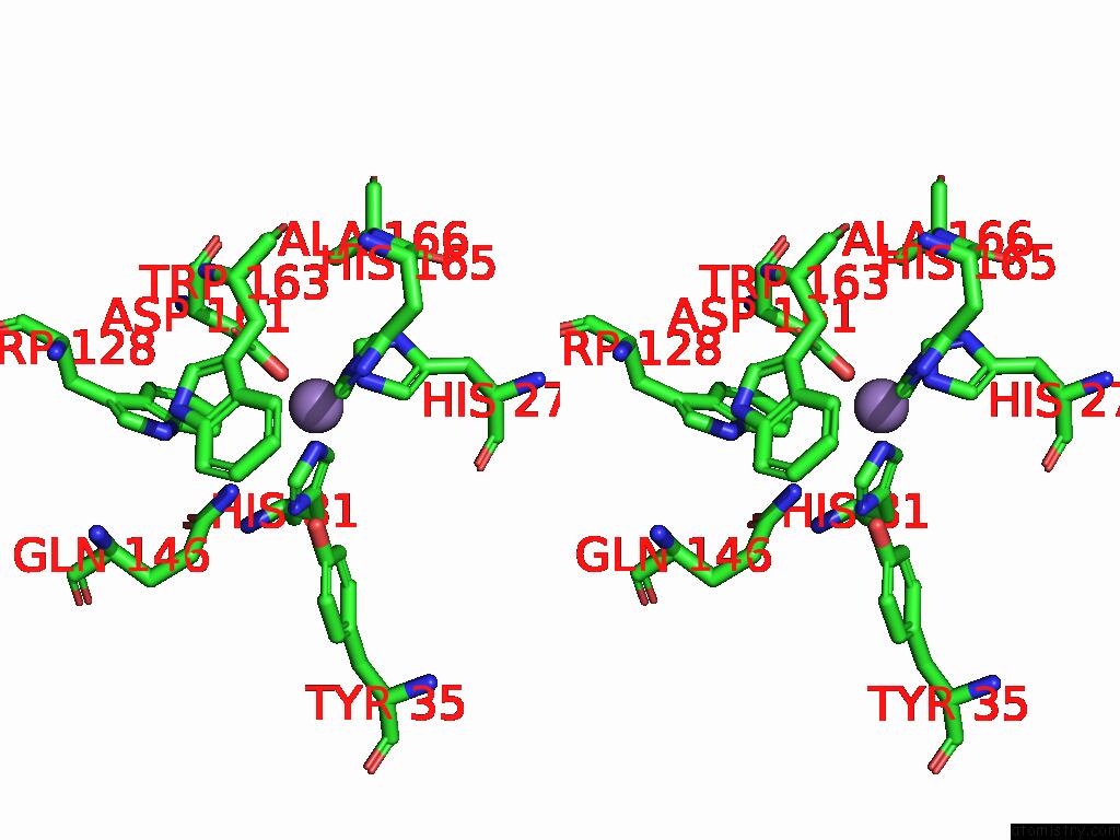
Stereo pair view
Mono view
Stereo pair view
A full contact list of Manganese with other atoms in the Mn binding
site number 2 of Crystal Structure of A Mutant Staphylococcus Equorum Manganese Superoxide Dismutase T89C and N187C within 5.0Å range:
|
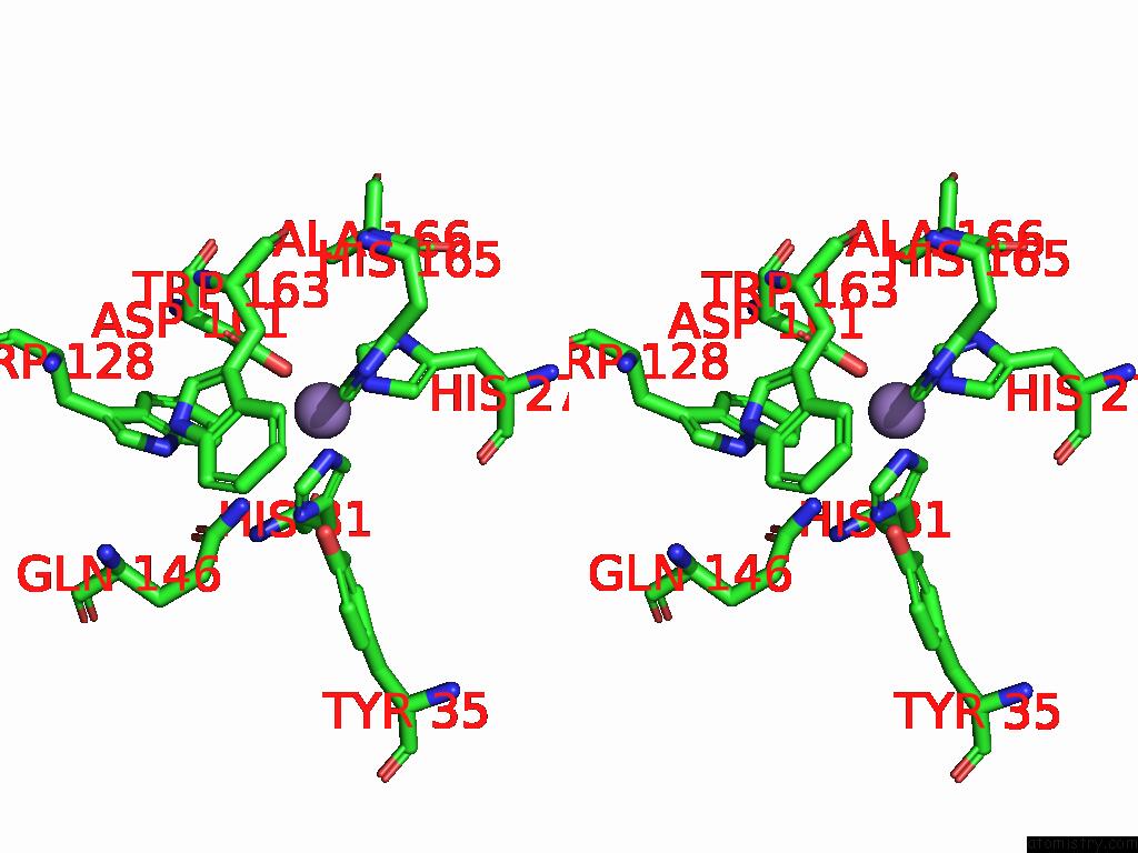
Manganese binding site 3 out of 4 in 8y17
Go back to
Manganese binding site 3 out
of 4 in the Crystal Structure of A Mutant Staphylococcus Equorum Manganese Superoxide Dismutase T89C and N187C

Mono view
Stereo pair view
Mono view
Stereo pair view
A full contact list of Manganese with other atoms in the Mn binding
site number 3 of Crystal Structure of A Mutant Staphylococcus Equorum Manganese Superoxide Dismutase T89C and N187C within 5.0Å range:
|
Manganese binding site 4 out of 4 in 8y17
Go back to
Manganese binding site 4 out
of 4 in the Crystal Structure of A Mutant Staphylococcus Equorum Manganese Superoxide Dismutase T89C and N187C
Mono view
Stereo pair view
Mono view
Stereo pair view
A full contact list of Manganese with other atoms in the Mn binding
site number 4 of Crystal Structure of A Mutant Staphylococcus Equorum Manganese Superoxide Dismutase T89C and N187C within 5.0Å range:
|
Reference:
N.A.Putri,
M.D.Fadillah,
H.Yoshida,
D.S.Retnoningrum,
A.A.Artarini,
R.A.Utami,
W.T.Ismaya.
Crystal Structure of A Mutant Staphylococcus Equorum Manganese Superoxide Dismutase T89C and N187C To Be Published.
Page generated: Sun Feb 9 08:27:43 2025
Last articles
Mg in 4PJ1Mg in 4PJJ
Mg in 4PJ3
Mg in 4PJ2
Mg in 4PIO
Mg in 4PIP
Mg in 4PHY
Mg in 4PHT
Mg in 4PHH
Mg in 4PHL