Manganese »
PDB 8sln-8uwb »
8ujf »
Manganese in PDB 8ujf: X-Ray Crystal Structure of Toxoplasma Gondii Galnac-T3 in Complex with Udp-Galnac, MN2+, and MUC5AC-3,13
Enzymatic activity of X-Ray Crystal Structure of Toxoplasma Gondii Galnac-T3 in Complex with Udp-Galnac, MN2+, and MUC5AC-3,13
All present enzymatic activity of X-Ray Crystal Structure of Toxoplasma Gondii Galnac-T3 in Complex with Udp-Galnac, MN2+, and MUC5AC-3,13:
2.4.1.41;
2.4.1.41;
Protein crystallography data
The structure of X-Ray Crystal Structure of Toxoplasma Gondii Galnac-T3 in Complex with Udp-Galnac, MN2+, and MUC5AC-3,13, PDB code: 8ujf
was solved by
P.Kumar,
N.L.Samara,
with X-Ray Crystallography technique. A brief refinement statistics is given in the table below:
| Resolution Low / High (Å) | 20.01 / 2.87 |
| Space group | P 21 21 21 |
| Cell size a, b, c (Å), α, β, γ (°) | 66.233, 123.693, 165.724, 90, 90, 90 |
| R / Rfree (%) | 21.7 / 27.4 |
Manganese Binding Sites:
The binding sites of Manganese atom in the X-Ray Crystal Structure of Toxoplasma Gondii Galnac-T3 in Complex with Udp-Galnac, MN2+, and MUC5AC-3,13
(pdb code 8ujf). This binding sites where shown within
5.0 Angstroms radius around Manganese atom.
In total 4 binding sites of Manganese where determined in the X-Ray Crystal Structure of Toxoplasma Gondii Galnac-T3 in Complex with Udp-Galnac, MN2+, and MUC5AC-3,13, PDB code: 8ujf:
Jump to Manganese binding site number: 1; 2; 3; 4;
In total 4 binding sites of Manganese where determined in the X-Ray Crystal Structure of Toxoplasma Gondii Galnac-T3 in Complex with Udp-Galnac, MN2+, and MUC5AC-3,13, PDB code: 8ujf:
Jump to Manganese binding site number: 1; 2; 3; 4;
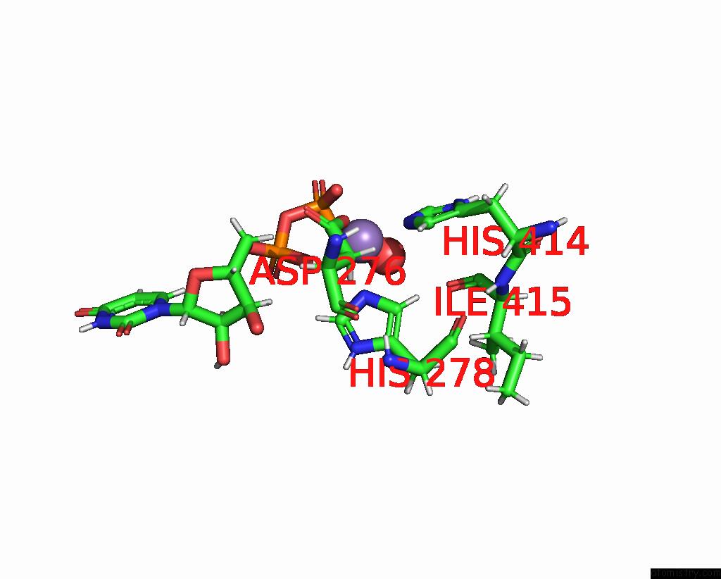
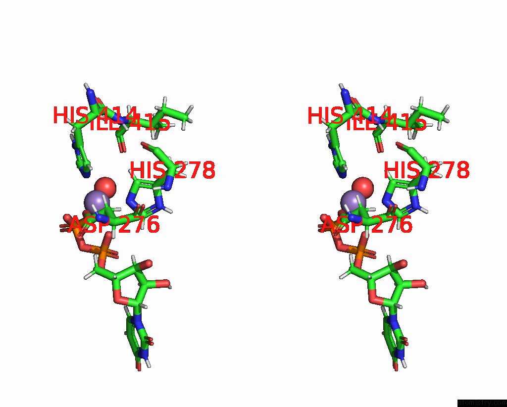
Manganese binding site 1 out of 4 in 8ujf
Go back to
Manganese binding site 1 out
of 4 in the X-Ray Crystal Structure of Toxoplasma Gondii Galnac-T3 in Complex with Udp-Galnac, MN2+, and MUC5AC-3,13
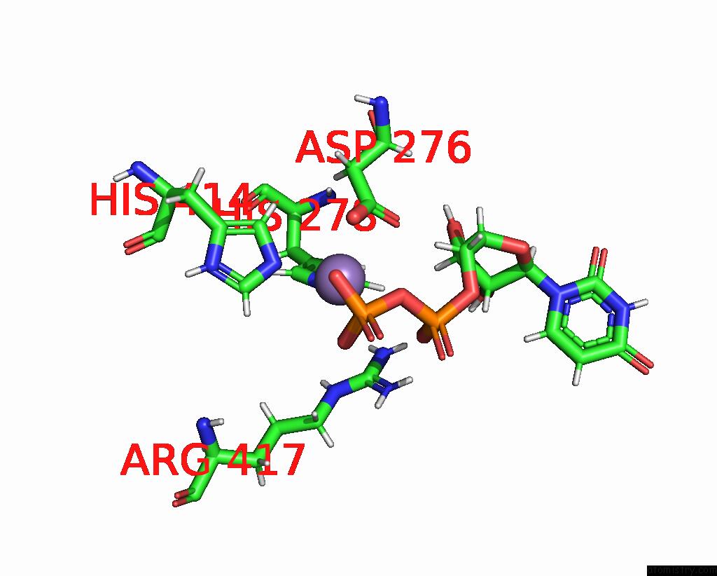
Mono view
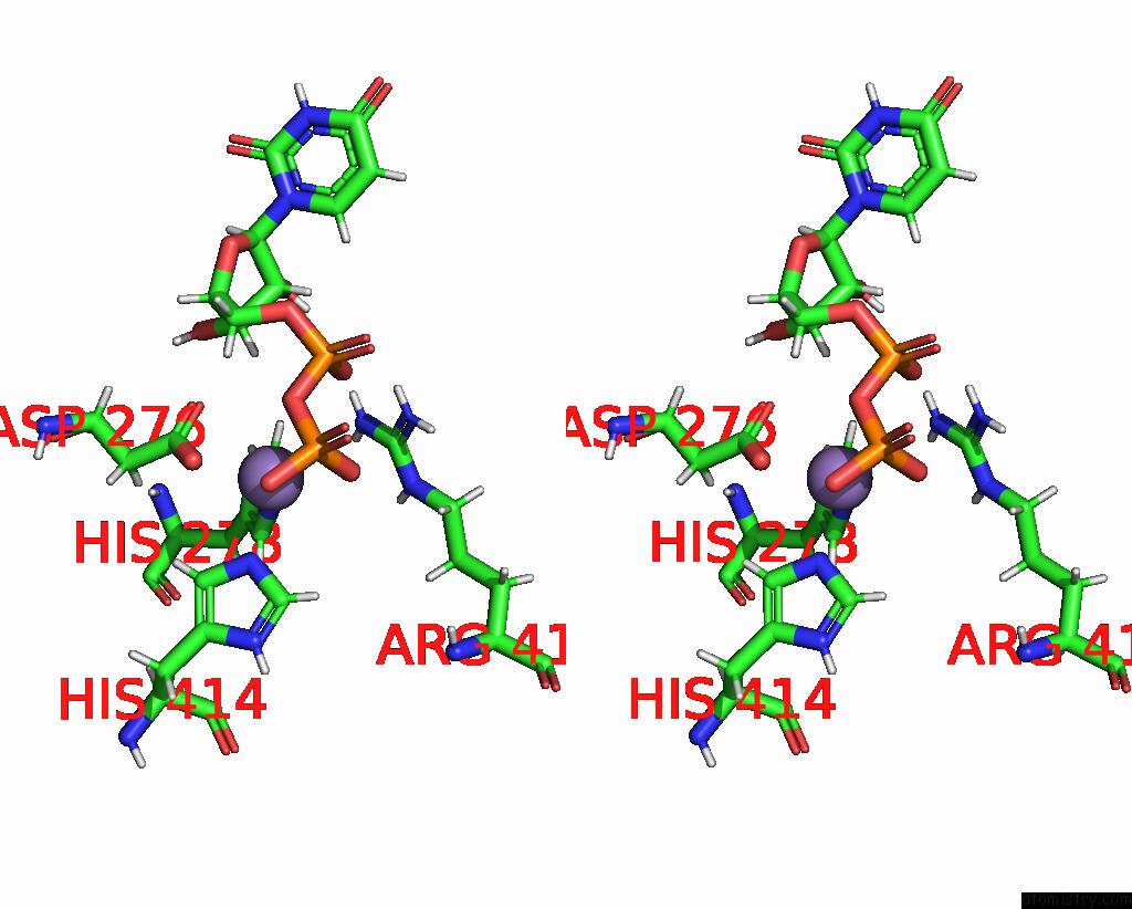
Stereo pair view
Mono view
Stereo pair view
A full contact list of Manganese with other atoms in the Mn binding
site number 1 of X-Ray Crystal Structure of Toxoplasma Gondii Galnac-T3 in Complex with Udp-Galnac, MN2+, and MUC5AC-3,13 within 5.0Å range:
|
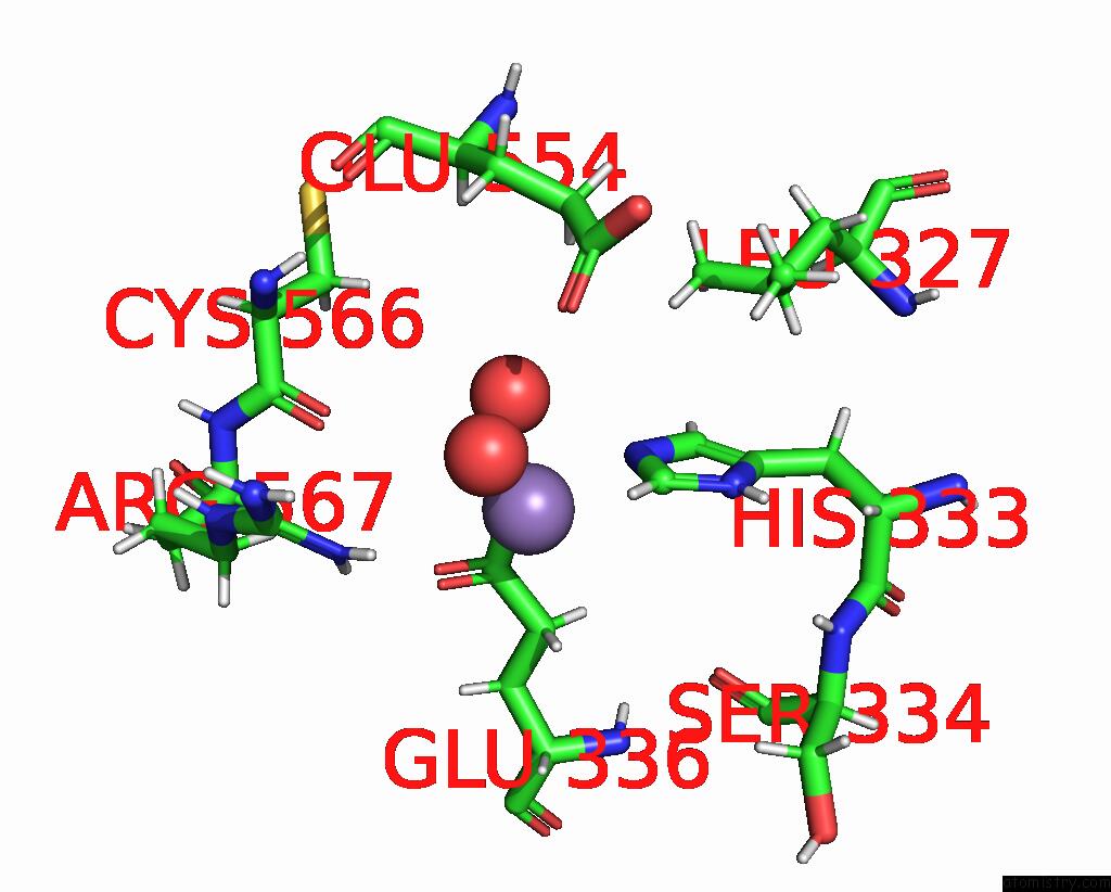
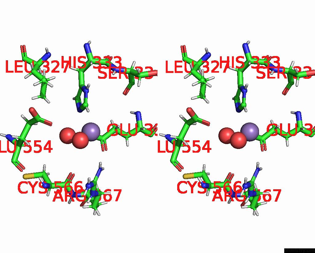
Manganese binding site 2 out of 4 in 8ujf
Go back to
Manganese binding site 2 out
of 4 in the X-Ray Crystal Structure of Toxoplasma Gondii Galnac-T3 in Complex with Udp-Galnac, MN2+, and MUC5AC-3,13
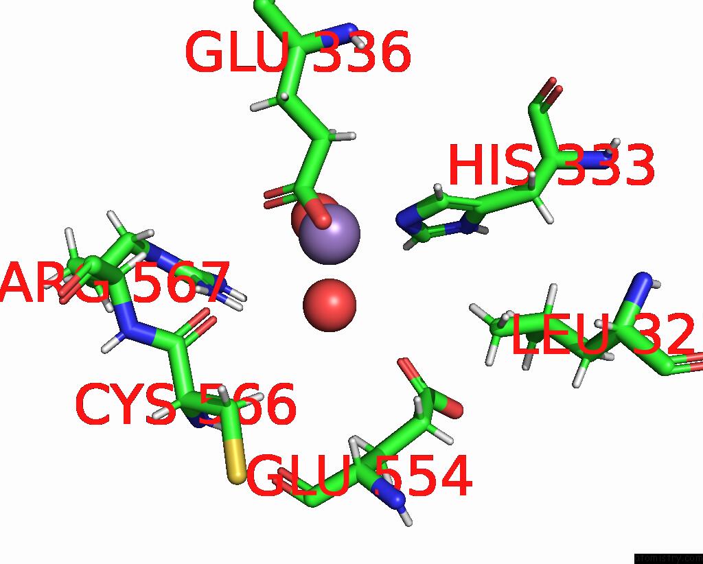
Mono view
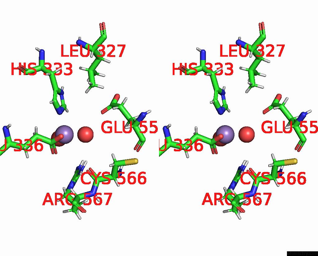
Stereo pair view
Mono view
Stereo pair view
A full contact list of Manganese with other atoms in the Mn binding
site number 2 of X-Ray Crystal Structure of Toxoplasma Gondii Galnac-T3 in Complex with Udp-Galnac, MN2+, and MUC5AC-3,13 within 5.0Å range:
|
Manganese binding site 3 out of 4 in 8ujf
Go back to
Manganese binding site 3 out
of 4 in the X-Ray Crystal Structure of Toxoplasma Gondii Galnac-T3 in Complex with Udp-Galnac, MN2+, and MUC5AC-3,13
Mono view
Stereo pair view
Mono view
Stereo pair view
A full contact list of Manganese with other atoms in the Mn binding
site number 3 of X-Ray Crystal Structure of Toxoplasma Gondii Galnac-T3 in Complex with Udp-Galnac, MN2+, and MUC5AC-3,13 within 5.0Å range:
|
Manganese binding site 4 out of 4 in 8ujf
Go back to
Manganese binding site 4 out
of 4 in the X-Ray Crystal Structure of Toxoplasma Gondii Galnac-T3 in Complex with Udp-Galnac, MN2+, and MUC5AC-3,13
Mono view
Stereo pair view
Mono view
Stereo pair view
A full contact list of Manganese with other atoms in the Mn binding
site number 4 of X-Ray Crystal Structure of Toxoplasma Gondii Galnac-T3 in Complex with Udp-Galnac, MN2+, and MUC5AC-3,13 within 5.0Å range:
|
Reference:
P.Kumar,
T.Tomita,
T.A.Gerken,
C.J.Ballard,
Y.S.Lee,
L.M.Weiss,
N.L.Samara.
A Toxoplasma Gondii O-Glycosyltransferase That Modulates Bradyzoite Cyst Wall Rigidity Is Distinct From Host Homologues Nat Commun V. 15 3792 2024.
ISSN: ESSN 2041-1723
DOI: 10.1038/S41467-024-48253-W
Page generated: Sun Oct 6 14:00:52 2024
ISSN: ESSN 2041-1723
DOI: 10.1038/S41467-024-48253-W
Last articles
K in 5FW5K in 5FQ5
K in 5FUE
K in 5FTB
K in 5FT0
K in 5FIY
K in 5FOG
K in 5FKT
K in 5FKS
K in 5FKQ