Manganese »
PDB 8kfu-8q1x »
8pzh »
Manganese in PDB 8pzh: Lpdd (H61A) Mutant
Protein crystallography data
The structure of Lpdd (H61A) Mutant, PDB code: 8pzh
was solved by
D.Gahloth,
D.Leys,
with X-Ray Crystallography technique. A brief refinement statistics is given in the table below:
| Resolution Low / High (Å) | 45.00 / 2.02 |
| Space group | I 2 2 2 |
| Cell size a, b, c (Å), α, β, γ (°) | 52.7, 91.58, 124.69, 90, 90, 90 |
| R / Rfree (%) | 21.8 / 24.6 |
Manganese Binding Sites:
The binding sites of Manganese atom in the Lpdd (H61A) Mutant
(pdb code 8pzh). This binding sites where shown within
5.0 Angstroms radius around Manganese atom.
In total only one binding site of Manganese was determined in the Lpdd (H61A) Mutant, PDB code: 8pzh:
In total only one binding site of Manganese was determined in the Lpdd (H61A) Mutant, PDB code: 8pzh:
Manganese binding site 1 out of 1 in 8pzh
Go back to
Manganese binding site 1 out
of 1 in the Lpdd (H61A) Mutant
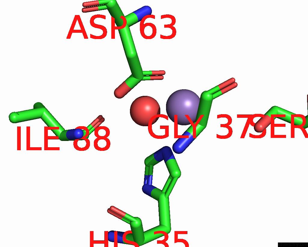
Mono view
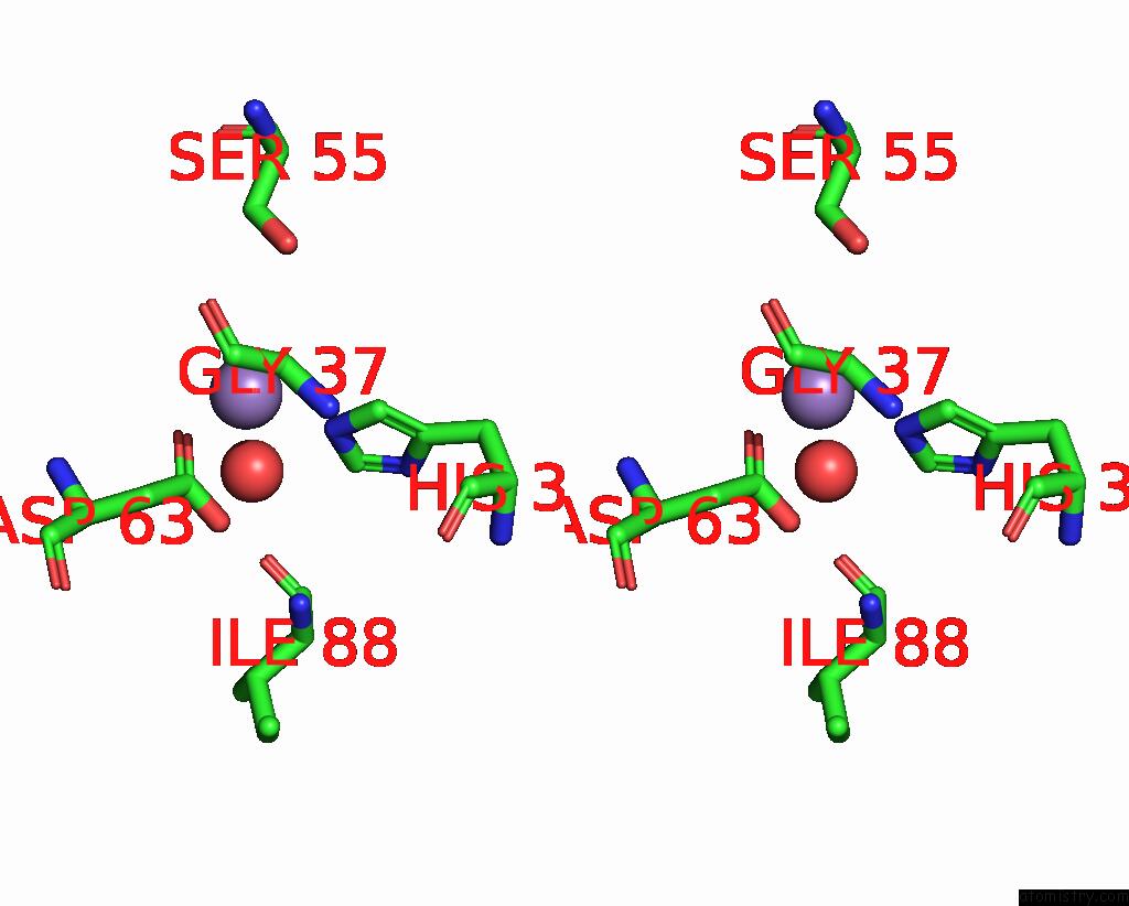
Stereo pair view
Mono view
Stereo pair view
A full contact list of Manganese with other atoms in the Mn binding
site number 1 of Lpdd (H61A) Mutant within 5.0Å range:
|
Reference:
D.Gahloth,
D.Leys.
Structure of Lpdd (H61A) Mutant From Lactobacillus Plantarum. To Be Published.
Page generated: Sun Oct 6 13:35:39 2024
Last articles
K in 4LWYK in 4LFC
K in 4LS5
K in 4LP8
K in 4LF6
K in 4LNG
K in 4LGN
K in 4LF8
K in 4LF7
K in 4LFA