Manganese »
PDB 8kfu-8q1x »
8oxg »
Manganese in PDB 8oxg: Crystal Structure of Human Methionine Aminopeptidase-2 Complexed with (3R,4S,5S,6R)-5-Methoxy-4-[(2R,3R)-2-Methyl-3-(3-Methyl-2-Buten-1- Yl)-2-Oxiranyl]-1-Oxaspiro[2.5]Oct-6-Yl N-(Trans-4-Aminocyclohexyl) Carbamate
Enzymatic activity of Crystal Structure of Human Methionine Aminopeptidase-2 Complexed with (3R,4S,5S,6R)-5-Methoxy-4-[(2R,3R)-2-Methyl-3-(3-Methyl-2-Buten-1- Yl)-2-Oxiranyl]-1-Oxaspiro[2.5]Oct-6-Yl N-(Trans-4-Aminocyclohexyl) Carbamate
All present enzymatic activity of Crystal Structure of Human Methionine Aminopeptidase-2 Complexed with (3R,4S,5S,6R)-5-Methoxy-4-[(2R,3R)-2-Methyl-3-(3-Methyl-2-Buten-1- Yl)-2-Oxiranyl]-1-Oxaspiro[2.5]Oct-6-Yl N-(Trans-4-Aminocyclohexyl) Carbamate:
3.4.11.18;
3.4.11.18;
Protein crystallography data
The structure of Crystal Structure of Human Methionine Aminopeptidase-2 Complexed with (3R,4S,5S,6R)-5-Methoxy-4-[(2R,3R)-2-Methyl-3-(3-Methyl-2-Buten-1- Yl)-2-Oxiranyl]-1-Oxaspiro[2.5]Oct-6-Yl N-(Trans-4-Aminocyclohexyl) Carbamate, PDB code: 8oxg
was solved by
S.Moss,
P.Cornelius,
with X-Ray Crystallography technique. A brief refinement statistics is given in the table below:
| Resolution Low / High (Å) | 44.84 / 1.73 |
| Space group | C 1 2 1 |
| Cell size a, b, c (Å), α, β, γ (°) | 119.05, 101.127, 83.737, 90, 98.76, 90 |
| R / Rfree (%) | 18.1 / 21.5 |
Manganese Binding Sites:
The binding sites of Manganese atom in the Crystal Structure of Human Methionine Aminopeptidase-2 Complexed with (3R,4S,5S,6R)-5-Methoxy-4-[(2R,3R)-2-Methyl-3-(3-Methyl-2-Buten-1- Yl)-2-Oxiranyl]-1-Oxaspiro[2.5]Oct-6-Yl N-(Trans-4-Aminocyclohexyl) Carbamate
(pdb code 8oxg). This binding sites where shown within
5.0 Angstroms radius around Manganese atom.
In total 4 binding sites of Manganese where determined in the Crystal Structure of Human Methionine Aminopeptidase-2 Complexed with (3R,4S,5S,6R)-5-Methoxy-4-[(2R,3R)-2-Methyl-3-(3-Methyl-2-Buten-1- Yl)-2-Oxiranyl]-1-Oxaspiro[2.5]Oct-6-Yl N-(Trans-4-Aminocyclohexyl) Carbamate, PDB code: 8oxg:
Jump to Manganese binding site number: 1; 2; 3; 4;
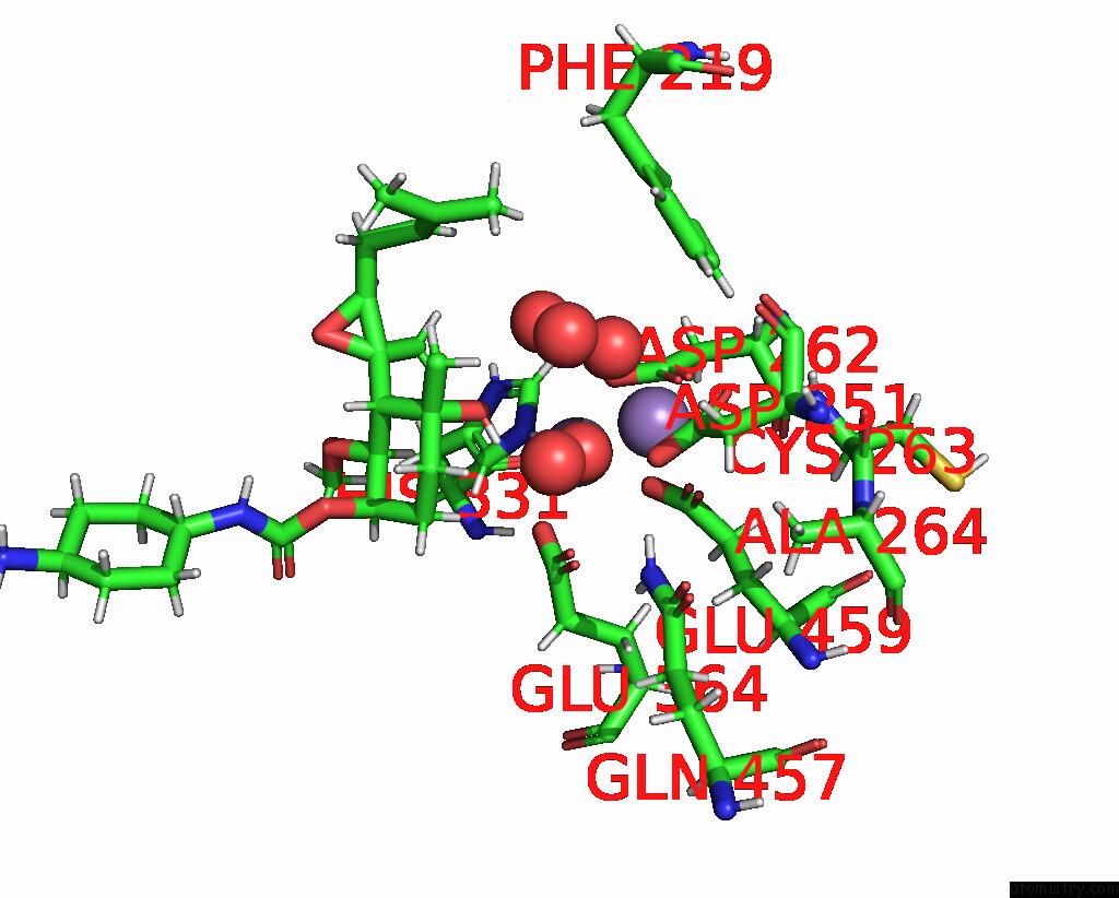
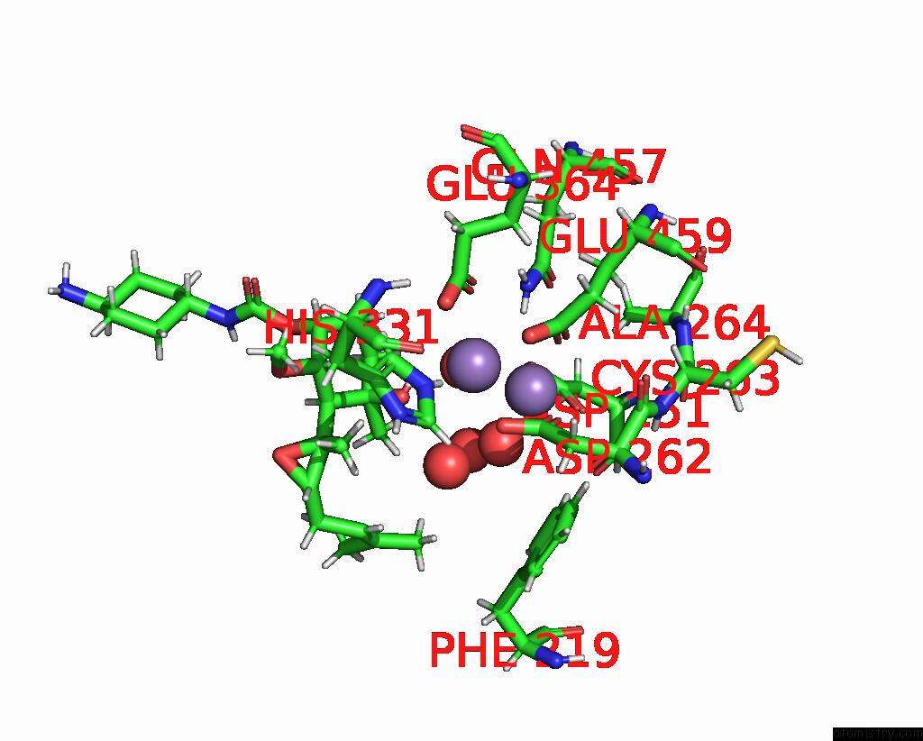
Manganese binding site 1 out of 4 in 8oxg
Go back to
Manganese binding site 1 out
of 4 in the Crystal Structure of Human Methionine Aminopeptidase-2 Complexed with (3R,4S,5S,6R)-5-Methoxy-4-[(2R,3R)-2-Methyl-3-(3-Methyl-2-Buten-1- Yl)-2-Oxiranyl]-1-Oxaspiro[2.5]Oct-6-Yl N-(Trans-4-Aminocyclohexyl) Carbamate
Mono view
Stereo pair view
Mono view
Stereo pair view
A full contact list of Manganese with other atoms in the Mn binding
site number 1 of Crystal Structure of Human Methionine Aminopeptidase-2 Complexed with (3R,4S,5S,6R)-5-Methoxy-4-[(2R,3R)-2-Methyl-3-(3-Methyl-2-Buten-1- Yl)-2-Oxiranyl]-1-Oxaspiro[2.5]Oct-6-Yl N-(Trans-4-Aminocyclohexyl) Carbamate within 5.0Å range:
|
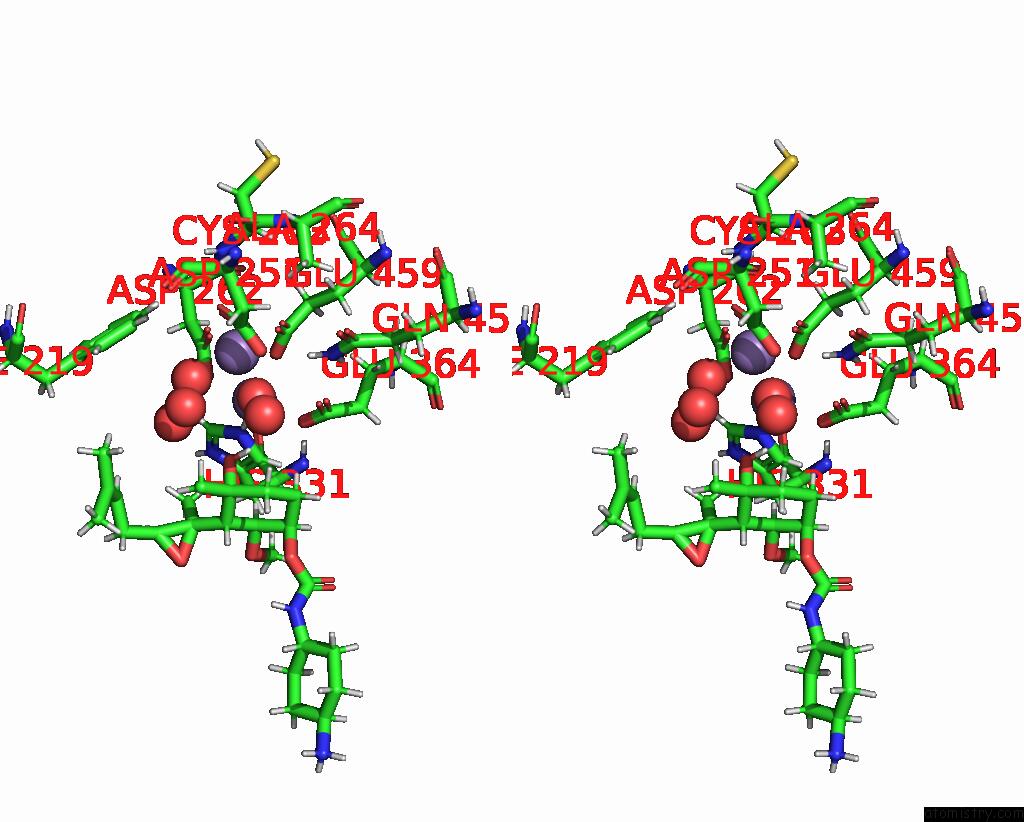
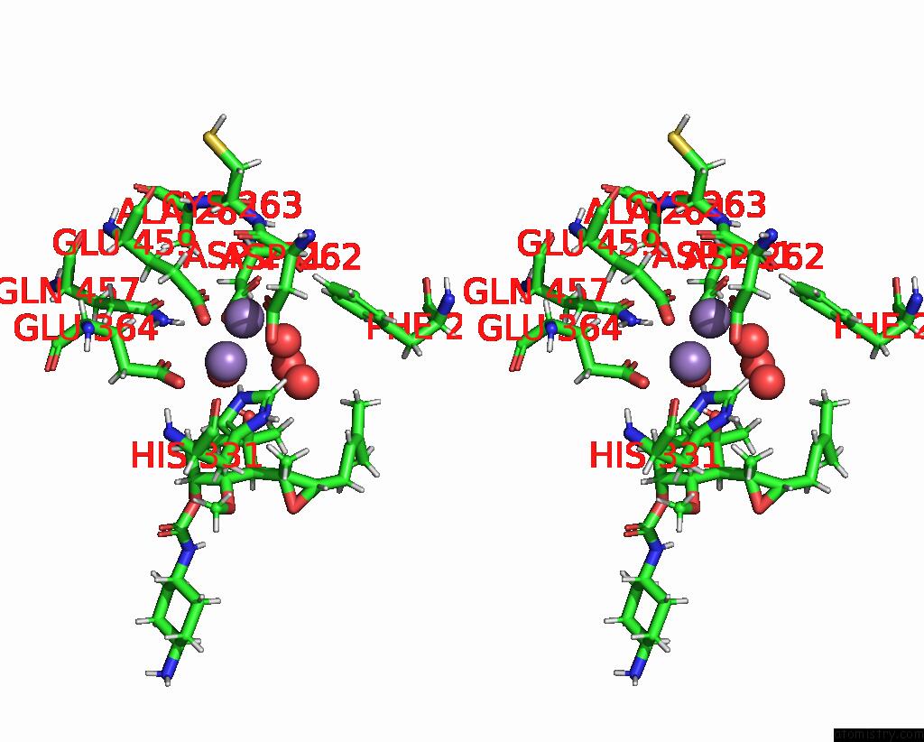
Manganese binding site 2 out of 4 in 8oxg
Go back to
Manganese binding site 2 out
of 4 in the Crystal Structure of Human Methionine Aminopeptidase-2 Complexed with (3R,4S,5S,6R)-5-Methoxy-4-[(2R,3R)-2-Methyl-3-(3-Methyl-2-Buten-1- Yl)-2-Oxiranyl]-1-Oxaspiro[2.5]Oct-6-Yl N-(Trans-4-Aminocyclohexyl) Carbamate
Mono view
Stereo pair view
Mono view
Stereo pair view
A full contact list of Manganese with other atoms in the Mn binding
site number 2 of Crystal Structure of Human Methionine Aminopeptidase-2 Complexed with (3R,4S,5S,6R)-5-Methoxy-4-[(2R,3R)-2-Methyl-3-(3-Methyl-2-Buten-1- Yl)-2-Oxiranyl]-1-Oxaspiro[2.5]Oct-6-Yl N-(Trans-4-Aminocyclohexyl) Carbamate within 5.0Å range:
|
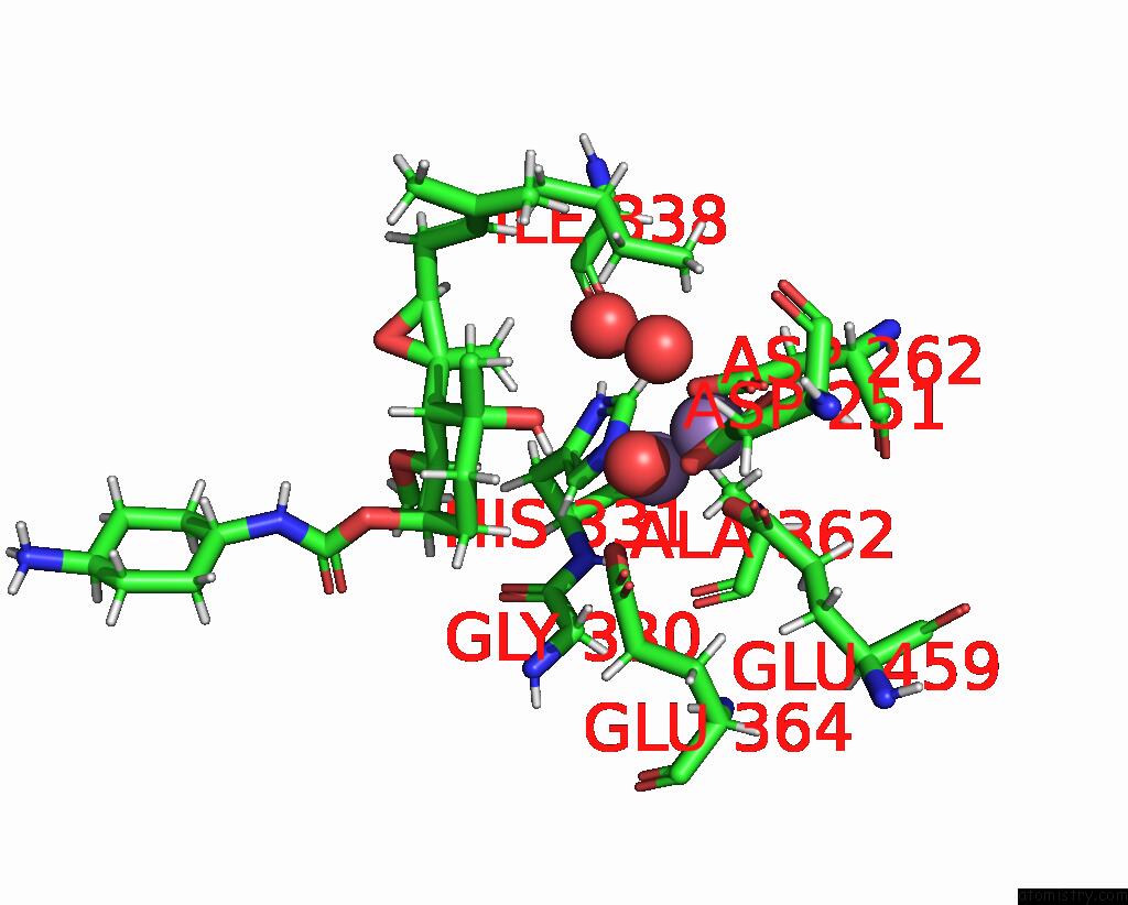
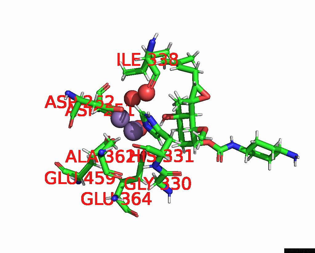
Manganese binding site 3 out of 4 in 8oxg
Go back to
Manganese binding site 3 out
of 4 in the Crystal Structure of Human Methionine Aminopeptidase-2 Complexed with (3R,4S,5S,6R)-5-Methoxy-4-[(2R,3R)-2-Methyl-3-(3-Methyl-2-Buten-1- Yl)-2-Oxiranyl]-1-Oxaspiro[2.5]Oct-6-Yl N-(Trans-4-Aminocyclohexyl) Carbamate
Mono view
Stereo pair view
Mono view
Stereo pair view
A full contact list of Manganese with other atoms in the Mn binding
site number 3 of Crystal Structure of Human Methionine Aminopeptidase-2 Complexed with (3R,4S,5S,6R)-5-Methoxy-4-[(2R,3R)-2-Methyl-3-(3-Methyl-2-Buten-1- Yl)-2-Oxiranyl]-1-Oxaspiro[2.5]Oct-6-Yl N-(Trans-4-Aminocyclohexyl) Carbamate within 5.0Å range:
|
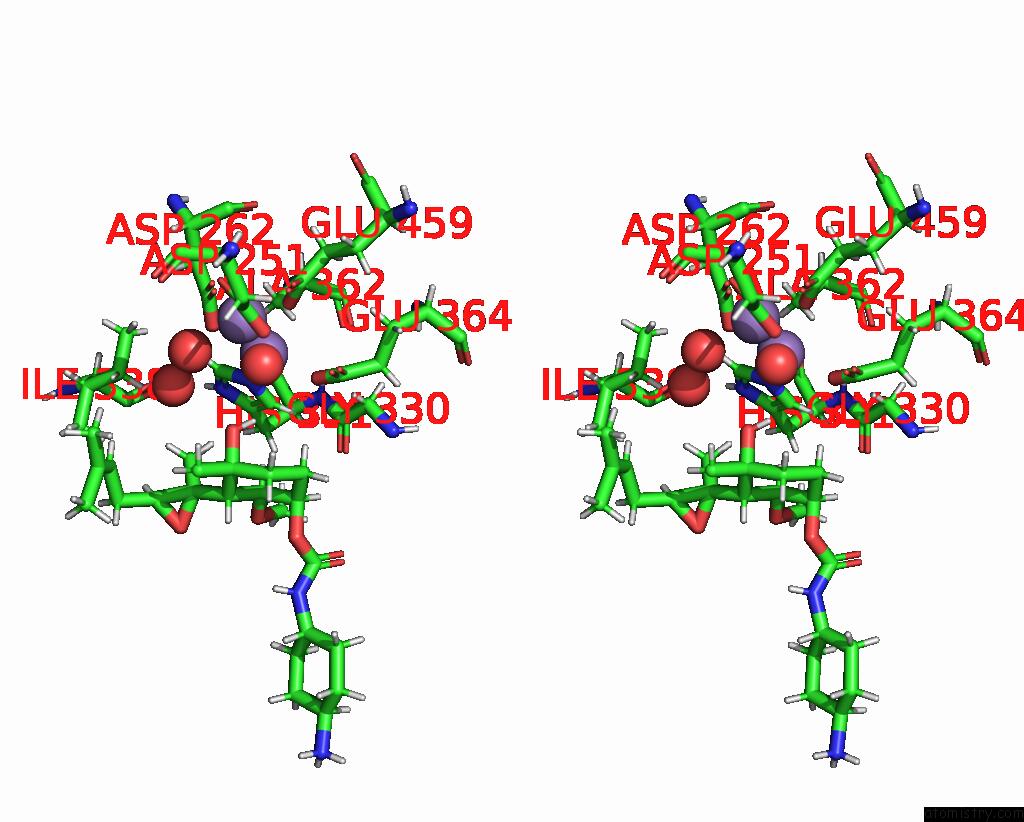
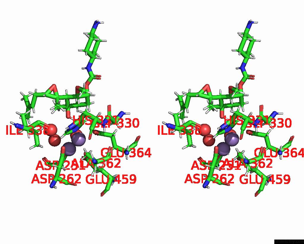
Manganese binding site 4 out of 4 in 8oxg
Go back to
Manganese binding site 4 out
of 4 in the Crystal Structure of Human Methionine Aminopeptidase-2 Complexed with (3R,4S,5S,6R)-5-Methoxy-4-[(2R,3R)-2-Methyl-3-(3-Methyl-2-Buten-1- Yl)-2-Oxiranyl]-1-Oxaspiro[2.5]Oct-6-Yl N-(Trans-4-Aminocyclohexyl) Carbamate
Mono view
Stereo pair view
Mono view
Stereo pair view
A full contact list of Manganese with other atoms in the Mn binding
site number 4 of Crystal Structure of Human Methionine Aminopeptidase-2 Complexed with (3R,4S,5S,6R)-5-Methoxy-4-[(2R,3R)-2-Methyl-3-(3-Methyl-2-Buten-1- Yl)-2-Oxiranyl]-1-Oxaspiro[2.5]Oct-6-Yl N-(Trans-4-Aminocyclohexyl) Carbamate within 5.0Å range:
|
Reference:
P.Cornelius,
B.A.Mayes,
J.S.Petersen,
D.J.Turnquist,
P.J.Dufour,
A.J.Dannenberg,
J.M.Shanahan,
B.J.Carver.
Pharmacological Characterization of Sdx-7320/Evexomostat: A Novel Methionine Aminopeptidase Type 2 Inhibitor with Anti-Tumor and Anti-Metastatic Activity. Mol.Cancer Ther. 2024.
ISSN: ESSN 1538-8514
PubMed: 38530115
DOI: 10.1158/1535-7163.MCT-23-0574
Page generated: Sun Oct 6 13:30:51 2024
ISSN: ESSN 1538-8514
PubMed: 38530115
DOI: 10.1158/1535-7163.MCT-23-0574
Last articles
K in 6CSRK in 6CSQ
K in 6CAO
K in 6CK4
K in 6CQ9
K in 6CSP
K in 6CQ8
K in 6CQ6
K in 6CP4
K in 6CNN