Manganese »
PDB 8dl1-8f4c »
8dq5 »
Manganese in PDB 8dq5: X-Ray Crystal Structure of Flavobacterium Johnsoniae Dimanganese(II) Class Id Ribonucleotide Reductase T191I Variant
Enzymatic activity of X-Ray Crystal Structure of Flavobacterium Johnsoniae Dimanganese(II) Class Id Ribonucleotide Reductase T191I Variant
All present enzymatic activity of X-Ray Crystal Structure of Flavobacterium Johnsoniae Dimanganese(II) Class Id Ribonucleotide Reductase T191I Variant:
1.17.4.1;
1.17.4.1;
Protein crystallography data
The structure of X-Ray Crystal Structure of Flavobacterium Johnsoniae Dimanganese(II) Class Id Ribonucleotide Reductase T191I Variant, PDB code: 8dq5
was solved by
H.R.Rose,
A.K.Boal,
with X-Ray Crystallography technique. A brief refinement statistics is given in the table below:
| Resolution Low / High (Å) | 46.57 / 2.10 |
| Space group | P 31 |
| Cell size a, b, c (Å), α, β, γ (°) | 53.771, 53.771, 221.288, 90, 90, 120 |
| R / Rfree (%) | 24.7 / 28.2 |
Other elements in 8dq5:
The structure of X-Ray Crystal Structure of Flavobacterium Johnsoniae Dimanganese(II) Class Id Ribonucleotide Reductase T191I Variant also contains other interesting chemical elements:
| Magnesium | (Mg) | 1 atom |
Manganese Binding Sites:
The binding sites of Manganese atom in the X-Ray Crystal Structure of Flavobacterium Johnsoniae Dimanganese(II) Class Id Ribonucleotide Reductase T191I Variant
(pdb code 8dq5). This binding sites where shown within
5.0 Angstroms radius around Manganese atom.
In total 4 binding sites of Manganese where determined in the X-Ray Crystal Structure of Flavobacterium Johnsoniae Dimanganese(II) Class Id Ribonucleotide Reductase T191I Variant, PDB code: 8dq5:
Jump to Manganese binding site number: 1; 2; 3; 4;
In total 4 binding sites of Manganese where determined in the X-Ray Crystal Structure of Flavobacterium Johnsoniae Dimanganese(II) Class Id Ribonucleotide Reductase T191I Variant, PDB code: 8dq5:
Jump to Manganese binding site number: 1; 2; 3; 4;
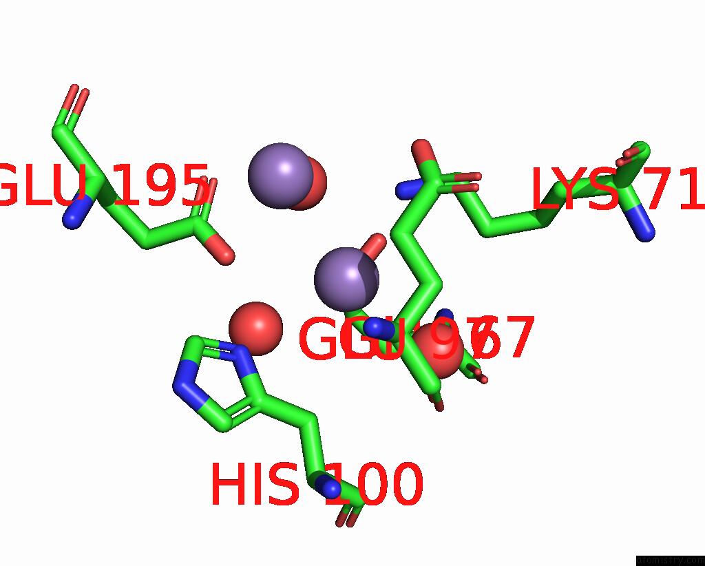
Manganese binding site 1 out of 4 in 8dq5
Go back to
Manganese binding site 1 out
of 4 in the X-Ray Crystal Structure of Flavobacterium Johnsoniae Dimanganese(II) Class Id Ribonucleotide Reductase T191I Variant

Mono view

Stereo pair view
Mono view
Stereo pair view
A full contact list of Manganese with other atoms in the Mn binding
site number 1 of X-Ray Crystal Structure of Flavobacterium Johnsoniae Dimanganese(II) Class Id Ribonucleotide Reductase T191I Variant within 5.0Å range:
|
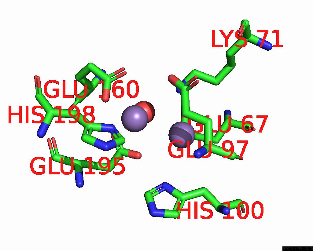
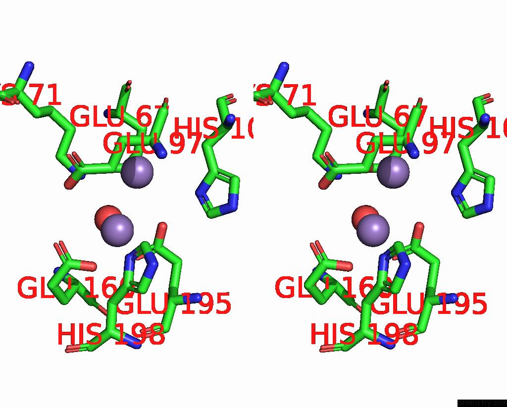
Manganese binding site 2 out of 4 in 8dq5
Go back to
Manganese binding site 2 out
of 4 in the X-Ray Crystal Structure of Flavobacterium Johnsoniae Dimanganese(II) Class Id Ribonucleotide Reductase T191I Variant

Mono view

Stereo pair view
Mono view
Stereo pair view
A full contact list of Manganese with other atoms in the Mn binding
site number 2 of X-Ray Crystal Structure of Flavobacterium Johnsoniae Dimanganese(II) Class Id Ribonucleotide Reductase T191I Variant within 5.0Å range:
|
Manganese binding site 3 out of 4 in 8dq5
Go back to
Manganese binding site 3 out
of 4 in the X-Ray Crystal Structure of Flavobacterium Johnsoniae Dimanganese(II) Class Id Ribonucleotide Reductase T191I Variant
Mono view

Stereo pair view
Mono view
Stereo pair view
A full contact list of Manganese with other atoms in the Mn binding
site number 3 of X-Ray Crystal Structure of Flavobacterium Johnsoniae Dimanganese(II) Class Id Ribonucleotide Reductase T191I Variant within 5.0Å range:
|
Manganese binding site 4 out of 4 in 8dq5
Go back to
Manganese binding site 4 out
of 4 in the X-Ray Crystal Structure of Flavobacterium Johnsoniae Dimanganese(II) Class Id Ribonucleotide Reductase T191I Variant
Mono view
Stereo pair view
Mono view
Stereo pair view
A full contact list of Manganese with other atoms in the Mn binding
site number 4 of X-Ray Crystal Structure of Flavobacterium Johnsoniae Dimanganese(II) Class Id Ribonucleotide Reductase T191I Variant within 5.0Å range:
|
Reference:
H.R.Rose,
A.K.Boal.
X-Ray Crystal Structure of Flavobacterium Johnsoniae Dimanganese(II) Class Id Ribonucleotide Reductase T191I Variant To Be Published.
Page generated: Sun Oct 6 11:33:37 2024
Last articles
K in 9JGZK in 9JH1
K in 9M3P
K in 9J0Z
K in 9J10
K in 9J3U
K in 9J0Y
K in 9J0X
K in 9ISK
K in 9I0M