Manganese »
PDB 8dl1-8f4c »
8dq3 »
Manganese in PDB 8dq3: X-Ray Crystal Structure of Aggregatibacter Actinomycetemcomitans Dimanganese(II) Class Id Ribonucleotide Reductase Beta Subunit
Enzymatic activity of X-Ray Crystal Structure of Aggregatibacter Actinomycetemcomitans Dimanganese(II) Class Id Ribonucleotide Reductase Beta Subunit
All present enzymatic activity of X-Ray Crystal Structure of Aggregatibacter Actinomycetemcomitans Dimanganese(II) Class Id Ribonucleotide Reductase Beta Subunit:
1.17.4.1;
1.17.4.1;
Protein crystallography data
The structure of X-Ray Crystal Structure of Aggregatibacter Actinomycetemcomitans Dimanganese(II) Class Id Ribonucleotide Reductase Beta Subunit, PDB code: 8dq3
was solved by
H.R.Rose,
J.J.Jung,
A.K.Boal,
with X-Ray Crystallography technique. A brief refinement statistics is given in the table below:
| Resolution Low / High (Å) | 44.22 / 1.67 |
| Space group | P 1 21 1 |
| Cell size a, b, c (Å), α, β, γ (°) | 52.213, 156.54, 80.722, 90, 91.09, 90 |
| R / Rfree (%) | 17 / 19.5 |
Other elements in 8dq3:
The structure of X-Ray Crystal Structure of Aggregatibacter Actinomycetemcomitans Dimanganese(II) Class Id Ribonucleotide Reductase Beta Subunit also contains other interesting chemical elements:
| Magnesium | (Mg) | 4 atoms |
Manganese Binding Sites:
The binding sites of Manganese atom in the X-Ray Crystal Structure of Aggregatibacter Actinomycetemcomitans Dimanganese(II) Class Id Ribonucleotide Reductase Beta Subunit
(pdb code 8dq3). This binding sites where shown within
5.0 Angstroms radius around Manganese atom.
In total 8 binding sites of Manganese where determined in the X-Ray Crystal Structure of Aggregatibacter Actinomycetemcomitans Dimanganese(II) Class Id Ribonucleotide Reductase Beta Subunit, PDB code: 8dq3:
Jump to Manganese binding site number: 1; 2; 3; 4; 5; 6; 7; 8;
In total 8 binding sites of Manganese where determined in the X-Ray Crystal Structure of Aggregatibacter Actinomycetemcomitans Dimanganese(II) Class Id Ribonucleotide Reductase Beta Subunit, PDB code: 8dq3:
Jump to Manganese binding site number: 1; 2; 3; 4; 5; 6; 7; 8;
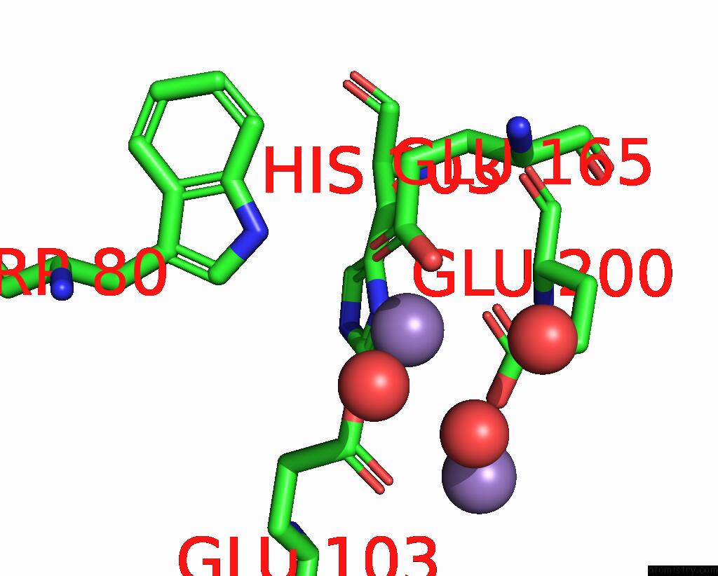
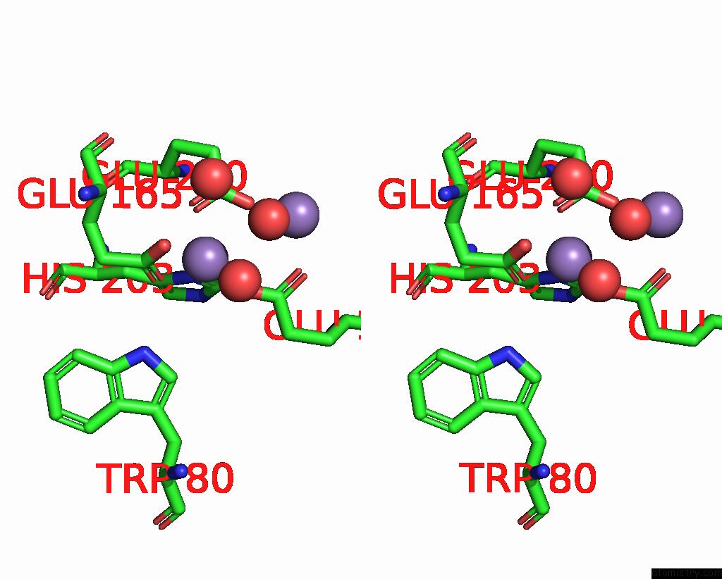
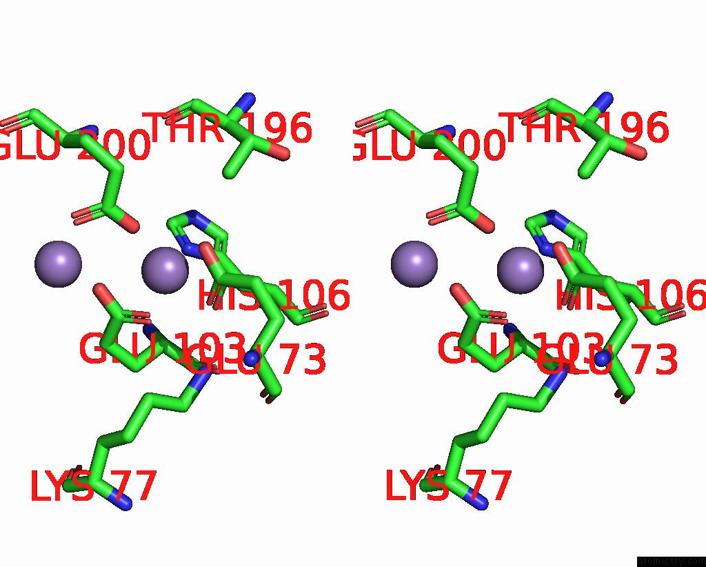
Manganese binding site 1 out of 8 in 8dq3
Go back to
Manganese binding site 1 out
of 8 in the X-Ray Crystal Structure of Aggregatibacter Actinomycetemcomitans Dimanganese(II) Class Id Ribonucleotide Reductase Beta Subunit
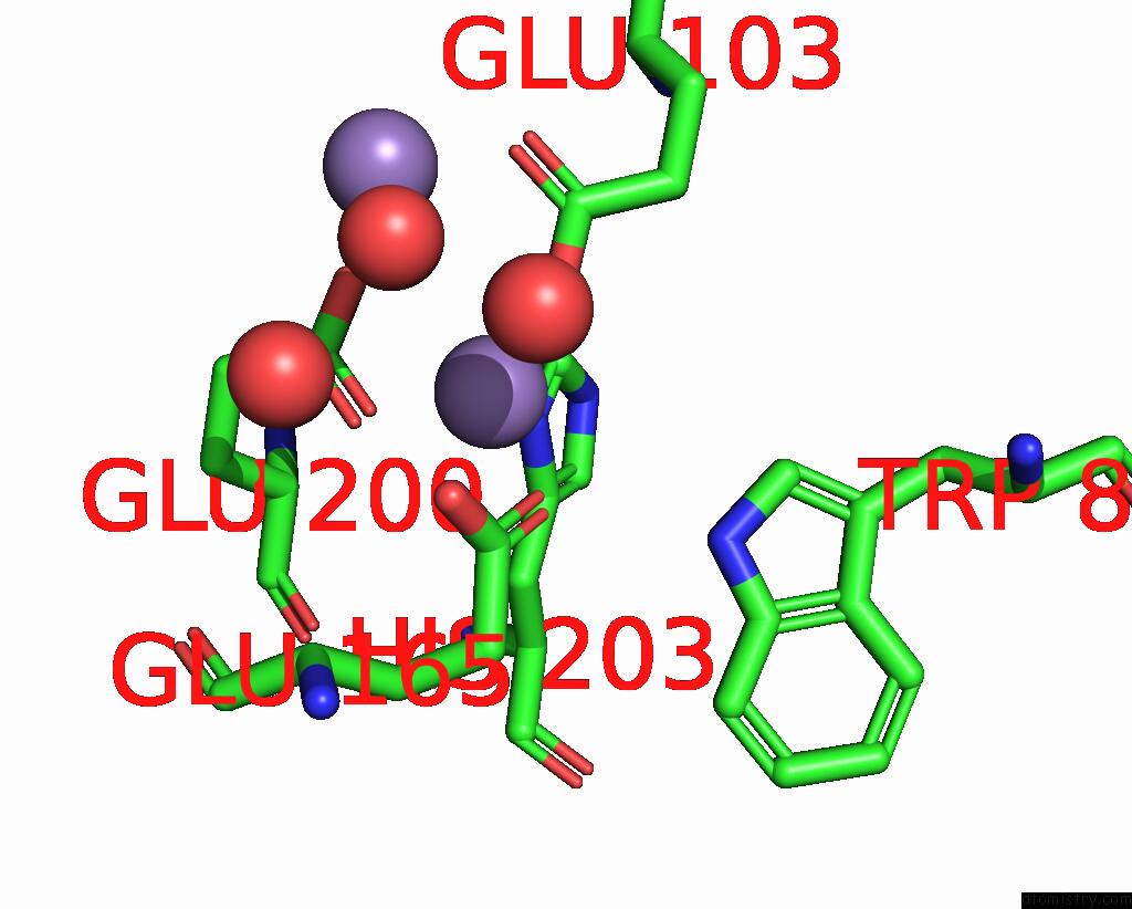
Mono view
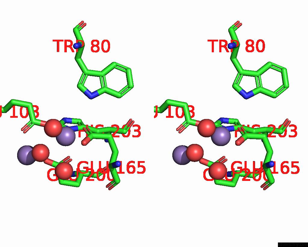
Stereo pair view
Mono view
Stereo pair view
A full contact list of Manganese with other atoms in the Mn binding
site number 1 of X-Ray Crystal Structure of Aggregatibacter Actinomycetemcomitans Dimanganese(II) Class Id Ribonucleotide Reductase Beta Subunit within 5.0Å range:
|
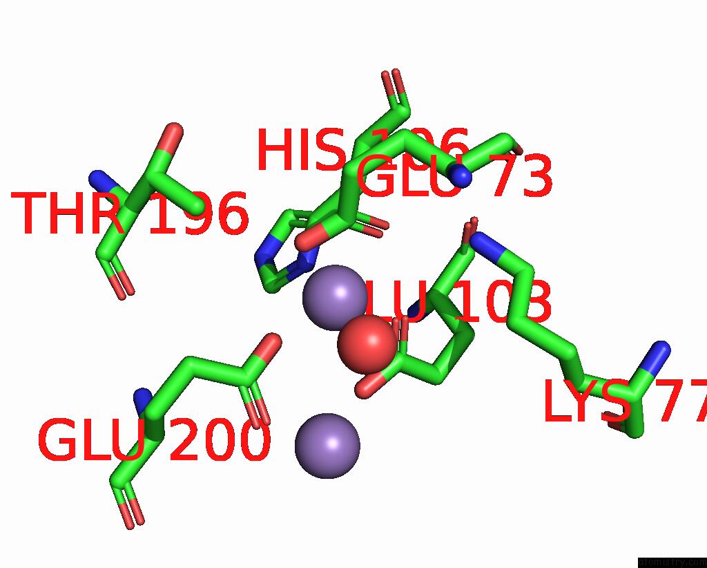
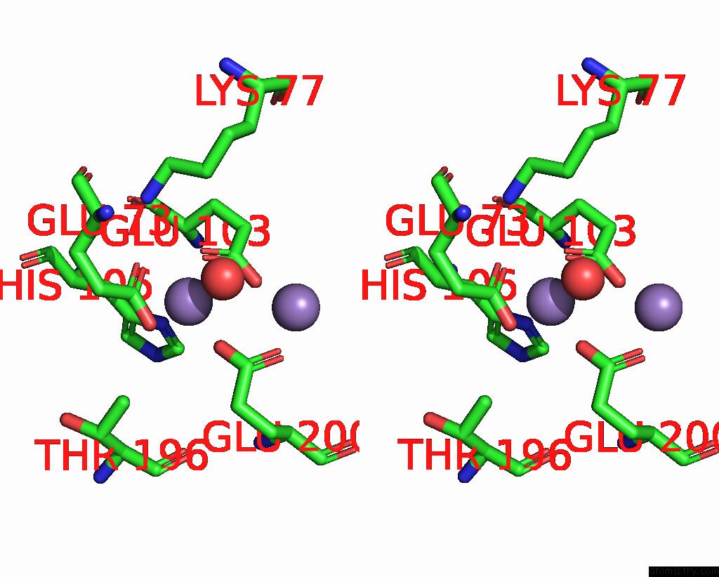
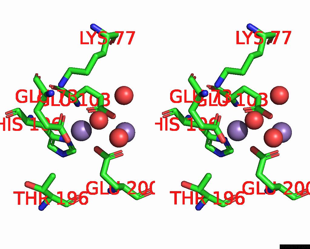
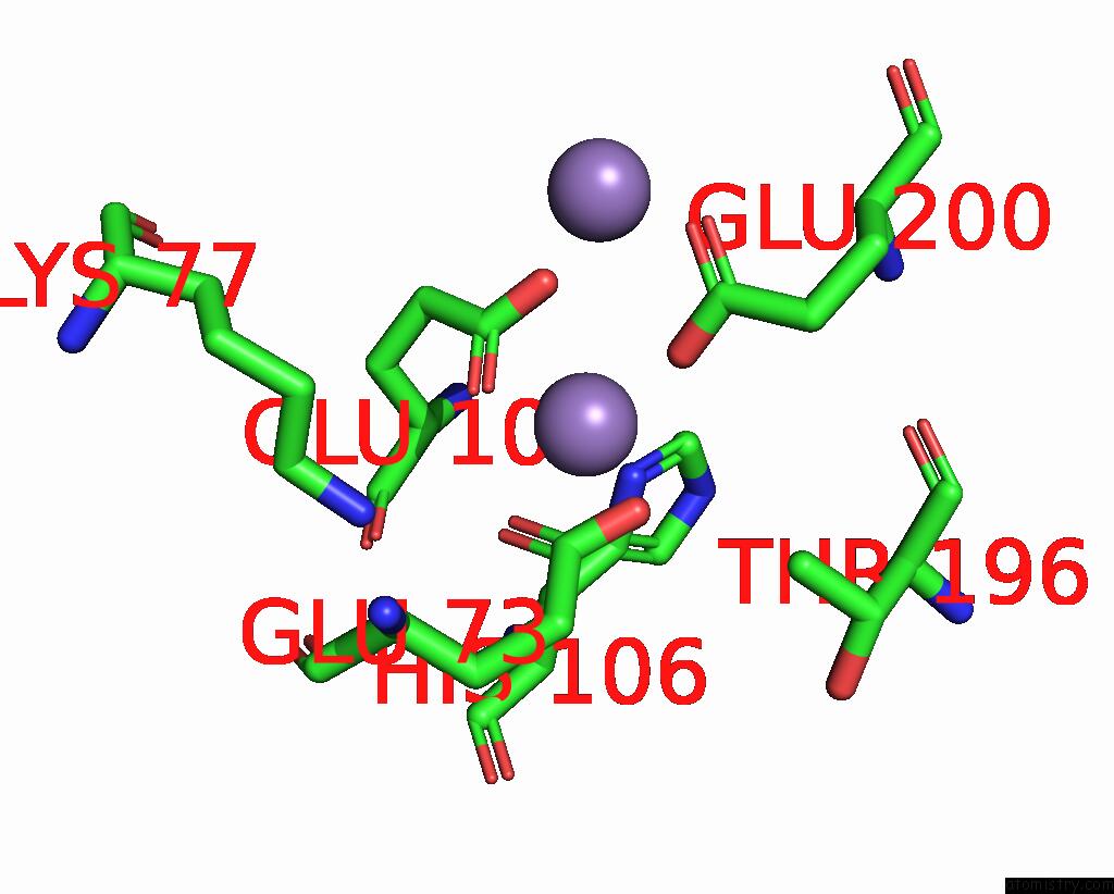
Manganese binding site 2 out of 8 in 8dq3
Go back to
Manganese binding site 2 out
of 8 in the X-Ray Crystal Structure of Aggregatibacter Actinomycetemcomitans Dimanganese(II) Class Id Ribonucleotide Reductase Beta Subunit

Mono view

Stereo pair view
Mono view
Stereo pair view
A full contact list of Manganese with other atoms in the Mn binding
site number 2 of X-Ray Crystal Structure of Aggregatibacter Actinomycetemcomitans Dimanganese(II) Class Id Ribonucleotide Reductase Beta Subunit within 5.0Å range:
|
Manganese binding site 3 out of 8 in 8dq3
Go back to
Manganese binding site 3 out
of 8 in the X-Ray Crystal Structure of Aggregatibacter Actinomycetemcomitans Dimanganese(II) Class Id Ribonucleotide Reductase Beta Subunit

Mono view

Stereo pair view
Mono view
Stereo pair view
A full contact list of Manganese with other atoms in the Mn binding
site number 3 of X-Ray Crystal Structure of Aggregatibacter Actinomycetemcomitans Dimanganese(II) Class Id Ribonucleotide Reductase Beta Subunit within 5.0Å range:
|
Manganese binding site 4 out of 8 in 8dq3
Go back to
Manganese binding site 4 out
of 8 in the X-Ray Crystal Structure of Aggregatibacter Actinomycetemcomitans Dimanganese(II) Class Id Ribonucleotide Reductase Beta Subunit
Mono view
Stereo pair view
Mono view
Stereo pair view
A full contact list of Manganese with other atoms in the Mn binding
site number 4 of X-Ray Crystal Structure of Aggregatibacter Actinomycetemcomitans Dimanganese(II) Class Id Ribonucleotide Reductase Beta Subunit within 5.0Å range:
|
Manganese binding site 5 out of 8 in 8dq3
Go back to
Manganese binding site 5 out
of 8 in the X-Ray Crystal Structure of Aggregatibacter Actinomycetemcomitans Dimanganese(II) Class Id Ribonucleotide Reductase Beta Subunit
Mono view
Stereo pair view
Mono view
Stereo pair view
A full contact list of Manganese with other atoms in the Mn binding
site number 5 of X-Ray Crystal Structure of Aggregatibacter Actinomycetemcomitans Dimanganese(II) Class Id Ribonucleotide Reductase Beta Subunit within 5.0Å range:
|
Manganese binding site 6 out of 8 in 8dq3
Go back to
Manganese binding site 6 out
of 8 in the X-Ray Crystal Structure of Aggregatibacter Actinomycetemcomitans Dimanganese(II) Class Id Ribonucleotide Reductase Beta Subunit

Mono view
Stereo pair view
Mono view
Stereo pair view
A full contact list of Manganese with other atoms in the Mn binding
site number 6 of X-Ray Crystal Structure of Aggregatibacter Actinomycetemcomitans Dimanganese(II) Class Id Ribonucleotide Reductase Beta Subunit within 5.0Å range:
|
Manganese binding site 7 out of 8 in 8dq3
Go back to
Manganese binding site 7 out
of 8 in the X-Ray Crystal Structure of Aggregatibacter Actinomycetemcomitans Dimanganese(II) Class Id Ribonucleotide Reductase Beta Subunit
Mono view
Stereo pair view
Mono view
Stereo pair view
A full contact list of Manganese with other atoms in the Mn binding
site number 7 of X-Ray Crystal Structure of Aggregatibacter Actinomycetemcomitans Dimanganese(II) Class Id Ribonucleotide Reductase Beta Subunit within 5.0Å range:
|
Manganese binding site 8 out of 8 in 8dq3
Go back to
Manganese binding site 8 out
of 8 in the X-Ray Crystal Structure of Aggregatibacter Actinomycetemcomitans Dimanganese(II) Class Id Ribonucleotide Reductase Beta Subunit

Mono view

Stereo pair view
Mono view
Stereo pair view
A full contact list of Manganese with other atoms in the Mn binding
site number 8 of X-Ray Crystal Structure of Aggregatibacter Actinomycetemcomitans Dimanganese(II) Class Id Ribonucleotide Reductase Beta Subunit within 5.0Å range:
|
Reference:
H.R.Rose,
A.K.Boal.
X-Ray Crystal Structure of Aggregatibacter Actinomycetemcomitans Dimanganese(II) Class Id Ribonucleotide Reductase Beta Subunit To Be Published.
Page generated: Sun Oct 6 11:33:38 2024
Last articles
Fe in 9KPPFe in 9KGP
Fe in 9K3V
Fe in 9K3U
Fe in 9KDG
Fe in 9KDF
Fe in 9K3B
Fe in 9K3Q
Fe in 9K39
Fe in 9K38