Manganese »
PDB 7x9j-7yzp »
7y1u »
Manganese in PDB 7y1u: Crystal Structure of Isocitrate Dehydrogenase From Campylobacter Corcagiensis
Enzymatic activity of Crystal Structure of Isocitrate Dehydrogenase From Campylobacter Corcagiensis
All present enzymatic activity of Crystal Structure of Isocitrate Dehydrogenase From Campylobacter Corcagiensis:
1.1.1.42;
1.1.1.42;
Protein crystallography data
The structure of Crystal Structure of Isocitrate Dehydrogenase From Campylobacter Corcagiensis, PDB code: 7y1u
was solved by
M.J.Bian,
Q.P.Cheng,
P.Wang,
G.P.Zhu,
with X-Ray Crystallography technique. A brief refinement statistics is given in the table below:
| Resolution Low / High (Å) | 48.93 / 2.50 |
| Space group | P 21 21 21 |
| Cell size a, b, c (Å), α, β, γ (°) | 62.501, 92.694, 147.321, 90, 90, 90 |
| R / Rfree (%) | 18.8 / 25.9 |
Manganese Binding Sites:
The binding sites of Manganese atom in the Crystal Structure of Isocitrate Dehydrogenase From Campylobacter Corcagiensis
(pdb code 7y1u). This binding sites where shown within
5.0 Angstroms radius around Manganese atom.
In total only one binding site of Manganese was determined in the Crystal Structure of Isocitrate Dehydrogenase From Campylobacter Corcagiensis, PDB code: 7y1u:
In total only one binding site of Manganese was determined in the Crystal Structure of Isocitrate Dehydrogenase From Campylobacter Corcagiensis, PDB code: 7y1u:
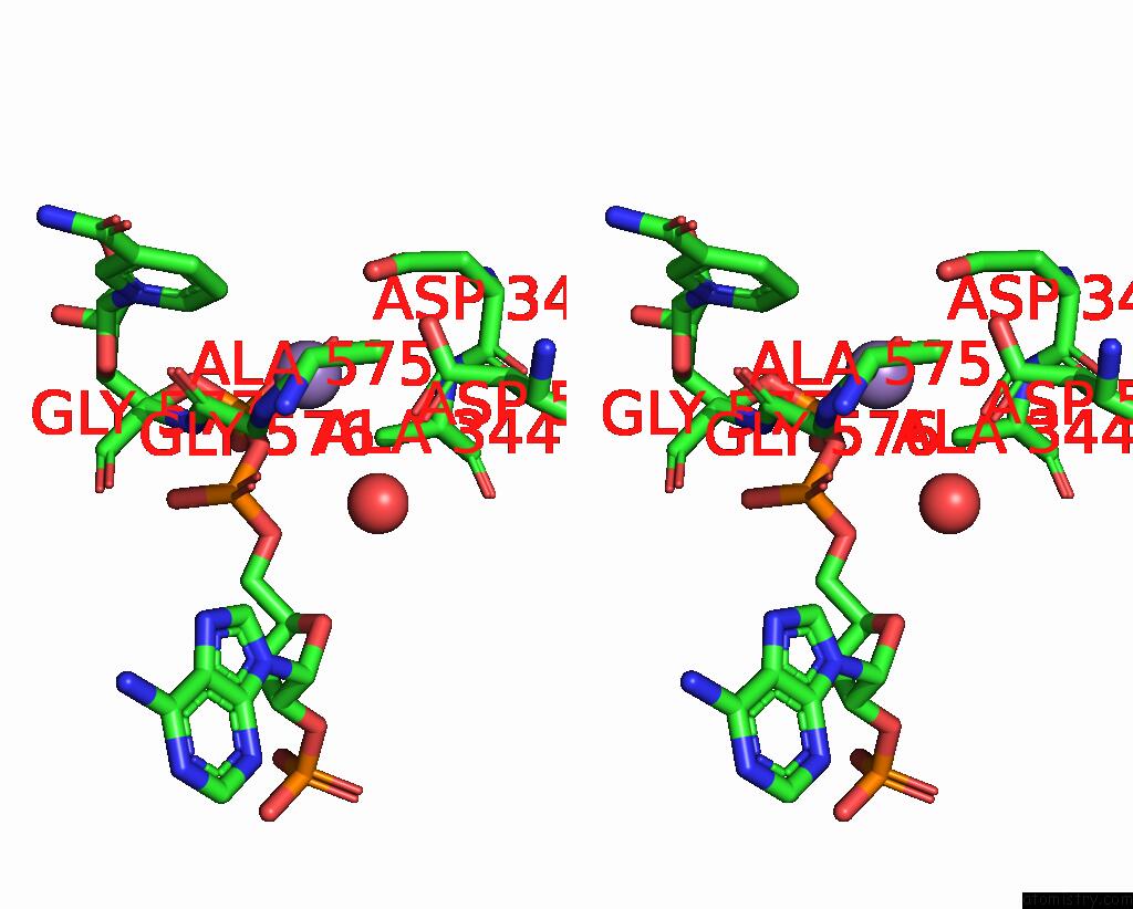
Manganese binding site 1 out of 1 in 7y1u
Go back to
Manganese binding site 1 out
of 1 in the Crystal Structure of Isocitrate Dehydrogenase From Campylobacter Corcagiensis

Mono view
Stereo pair view
Mono view
Stereo pair view
A full contact list of Manganese with other atoms in the Mn binding
site number 1 of Crystal Structure of Isocitrate Dehydrogenase From Campylobacter Corcagiensis within 5.0Å range:
|
Reference:
M.J.Bian,
Q.P.Cheng,
P.Wang,
G.P.Zhu.
Crystal Structure of Isocitrate Dehydrogenase From Campylobacter Corcagiensis To Be Published.
Page generated: Sun Aug 17 00:00:49 2025
Last articles
Ni in 5WTQNi in 5WXK
Ni in 5W1F
Ni in 5WWX
Ni in 5WRK
Ni in 5WK0
Ni in 5WAY
Ni in 5WJA
Ni in 5VMP
Ni in 5VGI