Manganese »
PDB 7uuh-7x9i »
7x9f »
Manganese in PDB 7x9f: Crystal Structure of Actinomycin D-Echinomycin-D(Agcgcgt/Acgcgct) Complex
Protein crystallography data
The structure of Crystal Structure of Actinomycin D-Echinomycin-D(Agcgcgt/Acgcgct) Complex, PDB code: 7x9f
was solved by
S.H.Kao,
R.B.Satange,
M.H.Hou,
with X-Ray Crystallography technique. A brief refinement statistics is given in the table below:
| Resolution Low / High (Å) | 27.80 / 2.96 |
| Space group | P 42 2 2 |
| Cell size a, b, c (Å), α, β, γ (°) | 87.917, 87.917, 47.551, 90, 90, 90 |
| R / Rfree (%) | 21.2 / 26.6 |
Manganese Binding Sites:
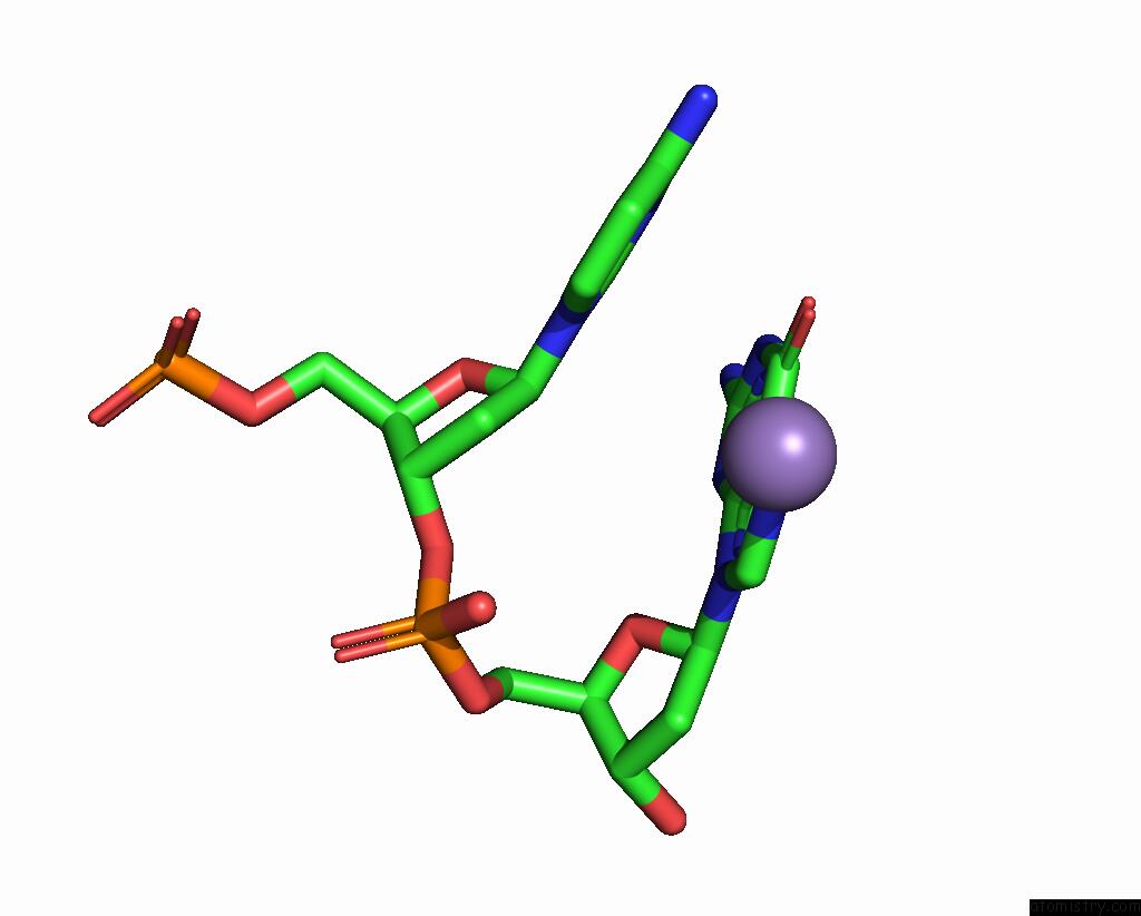
The binding sites of Manganese atom in the Crystal Structure of Actinomycin D-Echinomycin-D(Agcgcgt/Acgcgct) Complex
(pdb code 7x9f). This binding sites where shown within
5.0 Angstroms radius around Manganese atom.
In total only one binding site of Manganese was determined in the Crystal Structure of Actinomycin D-Echinomycin-D(Agcgcgt/Acgcgct) Complex, PDB code: 7x9f:
In total only one binding site of Manganese was determined in the Crystal Structure of Actinomycin D-Echinomycin-D(Agcgcgt/Acgcgct) Complex, PDB code: 7x9f:
Manganese binding site 1 out of 1 in 7x9f
Go back to
Manganese binding site 1 out
of 1 in the Crystal Structure of Actinomycin D-Echinomycin-D(Agcgcgt/Acgcgct) Complex
Mono view
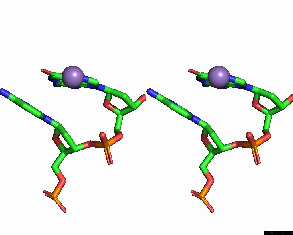
Stereo pair view
Mono view
Stereo pair view
A full contact list of Manganese with other atoms in the Mn binding
site number 1 of Crystal Structure of Actinomycin D-Echinomycin-D(Agcgcgt/Acgcgct) Complex within 5.0Å range:
|
Reference:
R.Satange,
S.H.Kao,
C.M.Chien,
S.H.Chou,
C.C.Lin,
S.Neidle,
M.H.Hou.
Staggered Intercalation of Dna Duplexes with Base-Pair Modulation By Two Distinct Drug Molecules Induces Asymmetric Backbone Twisting and Structure Polymorphism. Nucleic Acids Res. V. 50 8867 2022.
ISSN: ESSN 1362-4962
PubMed: 35871296
DOI: 10.1093/NAR/GKAC629
Page generated: Sat Aug 16 23:58:25 2025
ISSN: ESSN 1362-4962
PubMed: 35871296
DOI: 10.1093/NAR/GKAC629
Last articles
Ni in 5E6JNi in 5DQW
Ni in 5DQV
Ni in 5DOT
Ni in 5DMV
Ni in 5DMY
Ni in 5DFC
Ni in 5DFB
Ni in 5DAX
Ni in 5DAW