Manganese »
PDB 7uuh-7x9i »
7x7w »
Manganese in PDB 7x7w: The X-Ray Crystallographic Structure of D-Psicose 3-Epimerase From Clostridia Bacterium
Protein crystallography data
The structure of The X-Ray Crystallographic Structure of D-Psicose 3-Epimerase From Clostridia Bacterium, PDB code: 7x7w
was solved by
Z.F.Li,
X.F.Ban,
X.F.Xie,
Y.X.Tian,
C.M.Li,
Z.B.Gu,
with X-Ray Crystallography technique. A brief refinement statistics is given in the table below:
| Resolution Low / High (Å) | 37.94 / 2.10 |
| Space group | I 1 2 1 |
| Cell size a, b, c (Å), α, β, γ (°) | 60.018, 70.657, 153.784, 90, 99.32, 90 |
| R / Rfree (%) | 20 / 25.4 |
Manganese Binding Sites:
The binding sites of Manganese atom in the The X-Ray Crystallographic Structure of D-Psicose 3-Epimerase From Clostridia Bacterium
(pdb code 7x7w). This binding sites where shown within
5.0 Angstroms radius around Manganese atom.
In total 2 binding sites of Manganese where determined in the The X-Ray Crystallographic Structure of D-Psicose 3-Epimerase From Clostridia Bacterium, PDB code: 7x7w:
Jump to Manganese binding site number: 1; 2;
In total 2 binding sites of Manganese where determined in the The X-Ray Crystallographic Structure of D-Psicose 3-Epimerase From Clostridia Bacterium, PDB code: 7x7w:
Jump to Manganese binding site number: 1; 2;
Manganese binding site 1 out of 2 in 7x7w
Go back to
Manganese binding site 1 out
of 2 in the The X-Ray Crystallographic Structure of D-Psicose 3-Epimerase From Clostridia Bacterium
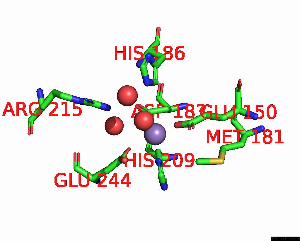
Mono view
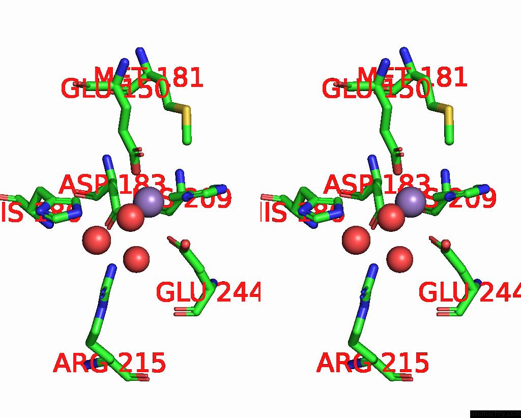
Stereo pair view
Mono view
Stereo pair view
A full contact list of Manganese with other atoms in the Mn binding
site number 1 of The X-Ray Crystallographic Structure of D-Psicose 3-Epimerase From Clostridia Bacterium within 5.0Å range:
|
Manganese binding site 2 out of 2 in 7x7w
Go back to
Manganese binding site 2 out
of 2 in the The X-Ray Crystallographic Structure of D-Psicose 3-Epimerase From Clostridia Bacterium
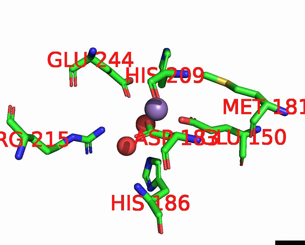
Mono view
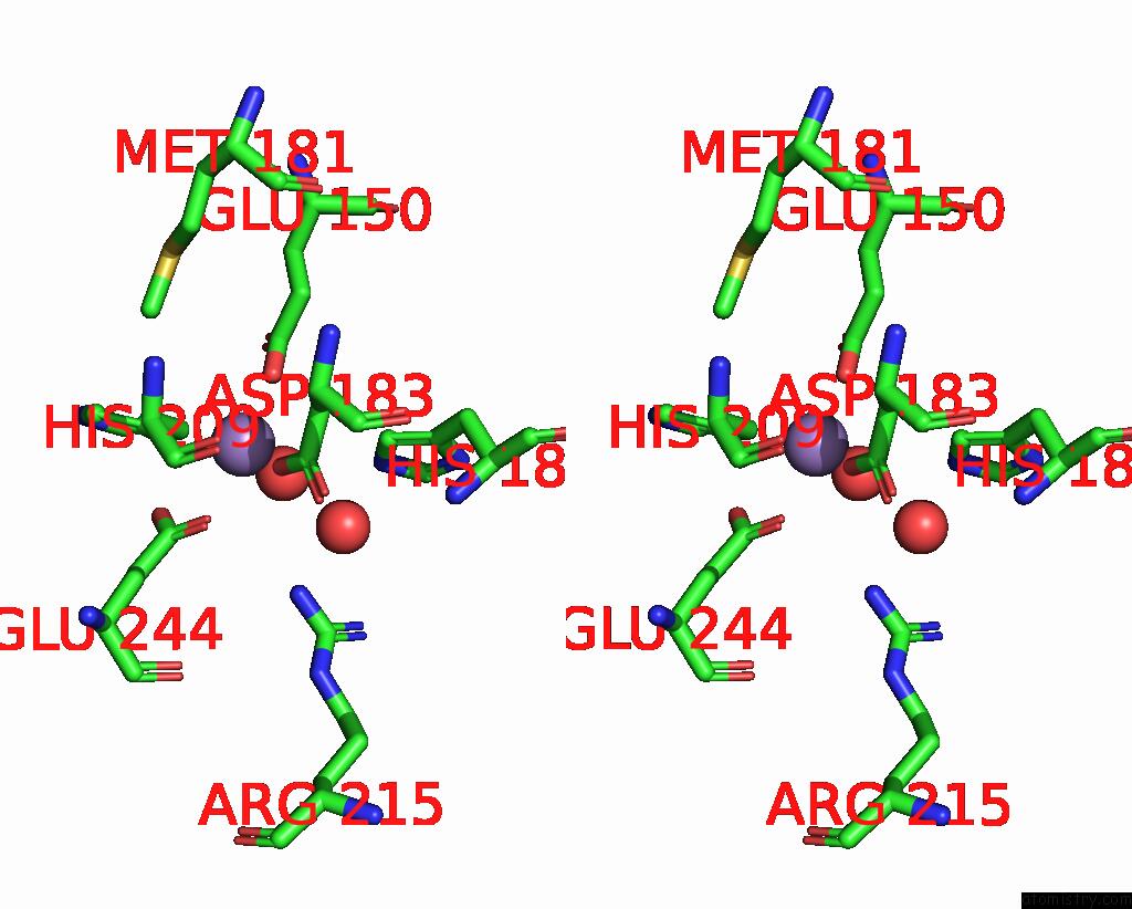
Stereo pair view
Mono view
Stereo pair view
A full contact list of Manganese with other atoms in the Mn binding
site number 2 of The X-Ray Crystallographic Structure of D-Psicose 3-Epimerase From Clostridia Bacterium within 5.0Å range:
|
Reference:
X.Xie,
Y.Tian,
X.Ban,
C.Li,
H.Yang,
Z.Li.
Crystal Structure of A Novel Homodimeric D-Allulose 3-Epimerase From A Clostridia Bacterium Acta Crystallogr.,Sect.D V. 78 1180 2022.
ISSN: ESSN 1399-0047
DOI: 10.1107/S2059798322007707
Page generated: Sat Aug 16 23:58:22 2025
ISSN: ESSN 1399-0047
DOI: 10.1107/S2059798322007707
Last articles
Ni in 5FPZNi in 5FPX
Ni in 5FM8
Ni in 5FLH
Ni in 5FLE
Ni in 5FJH
Ni in 5FJK
Ni in 5F5I
Ni in 5ERL
Ni in 5FG6