Manganese »
PDB 7uuh-7x9i »
7wnk »
Manganese in PDB 7wnk: Crystal Structure of A Mutant Staphylococcus Equorum Manganese Superoxide Dismutase K38R and A121E
Enzymatic activity of Crystal Structure of A Mutant Staphylococcus Equorum Manganese Superoxide Dismutase K38R and A121E
All present enzymatic activity of Crystal Structure of A Mutant Staphylococcus Equorum Manganese Superoxide Dismutase K38R and A121E:
1.15.1.1;
1.15.1.1;
Protein crystallography data
The structure of Crystal Structure of A Mutant Staphylococcus Equorum Manganese Superoxide Dismutase K38R and A121E, PDB code: 7wnk
was solved by
D.S.Retnoningrum,
H.Yoshida,
A.A.Artarini,
W.T.Ismaya,
with X-Ray Crystallography technique. A brief refinement statistics is given in the table below:
| Resolution Low / High (Å) | 44.94 / 1.40 |
| Space group | P 21 21 21 |
| Cell size a, b, c (Å), α, β, γ (°) | 73.765, 113.174, 119.908, 90, 90, 90 |
| R / Rfree (%) | 17.8 / 19.4 |
Other elements in 7wnk:
The structure of Crystal Structure of A Mutant Staphylococcus Equorum Manganese Superoxide Dismutase K38R and A121E also contains other interesting chemical elements:
| Bromine | (Br) | 1 atom |
Manganese Binding Sites:
The binding sites of Manganese atom in the Crystal Structure of A Mutant Staphylococcus Equorum Manganese Superoxide Dismutase K38R and A121E
(pdb code 7wnk). This binding sites where shown within
5.0 Angstroms radius around Manganese atom.
In total 4 binding sites of Manganese where determined in the Crystal Structure of A Mutant Staphylococcus Equorum Manganese Superoxide Dismutase K38R and A121E, PDB code: 7wnk:
Jump to Manganese binding site number: 1; 2; 3; 4;
In total 4 binding sites of Manganese where determined in the Crystal Structure of A Mutant Staphylococcus Equorum Manganese Superoxide Dismutase K38R and A121E, PDB code: 7wnk:
Jump to Manganese binding site number: 1; 2; 3; 4;
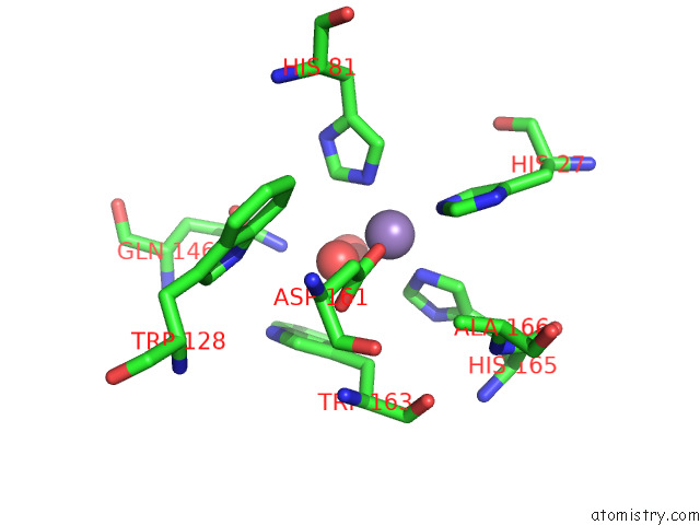
Manganese binding site 1 out of 4 in 7wnk
Go back to
Manganese binding site 1 out
of 4 in the Crystal Structure of A Mutant Staphylococcus Equorum Manganese Superoxide Dismutase K38R and A121E
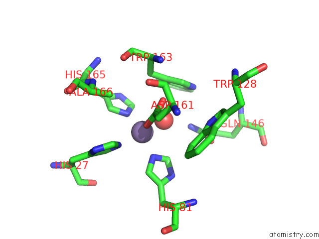
Mono view
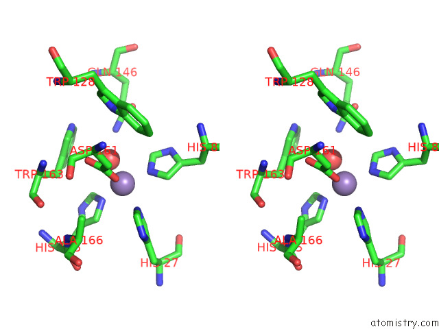
Stereo pair view
Mono view
Stereo pair view
A full contact list of Manganese with other atoms in the Mn binding
site number 1 of Crystal Structure of A Mutant Staphylococcus Equorum Manganese Superoxide Dismutase K38R and A121E within 5.0Å range:
|
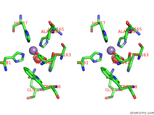
Manganese binding site 2 out of 4 in 7wnk
Go back to
Manganese binding site 2 out
of 4 in the Crystal Structure of A Mutant Staphylococcus Equorum Manganese Superoxide Dismutase K38R and A121E
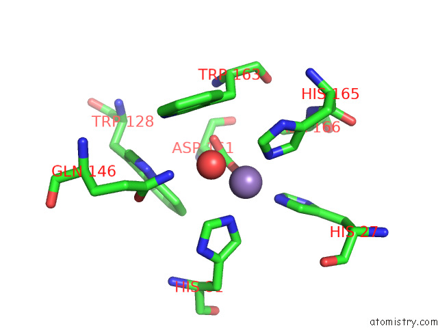
Mono view
Stereo pair view
Mono view
Stereo pair view
A full contact list of Manganese with other atoms in the Mn binding
site number 2 of Crystal Structure of A Mutant Staphylococcus Equorum Manganese Superoxide Dismutase K38R and A121E within 5.0Å range:
|
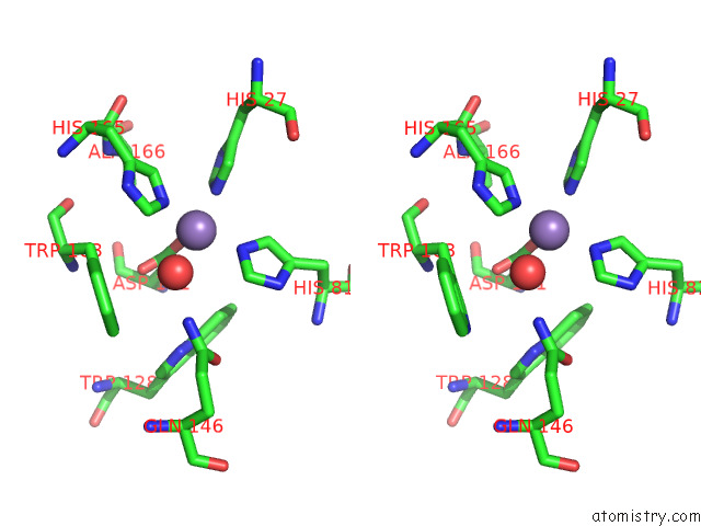
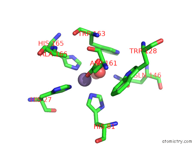
Manganese binding site 3 out of 4 in 7wnk
Go back to
Manganese binding site 3 out
of 4 in the Crystal Structure of A Mutant Staphylococcus Equorum Manganese Superoxide Dismutase K38R and A121E
Mono view
Stereo pair view
Mono view
Stereo pair view
A full contact list of Manganese with other atoms in the Mn binding
site number 3 of Crystal Structure of A Mutant Staphylococcus Equorum Manganese Superoxide Dismutase K38R and A121E within 5.0Å range:
|
Manganese binding site 4 out of 4 in 7wnk
Go back to
Manganese binding site 4 out
of 4 in the Crystal Structure of A Mutant Staphylococcus Equorum Manganese Superoxide Dismutase K38R and A121E
Mono view
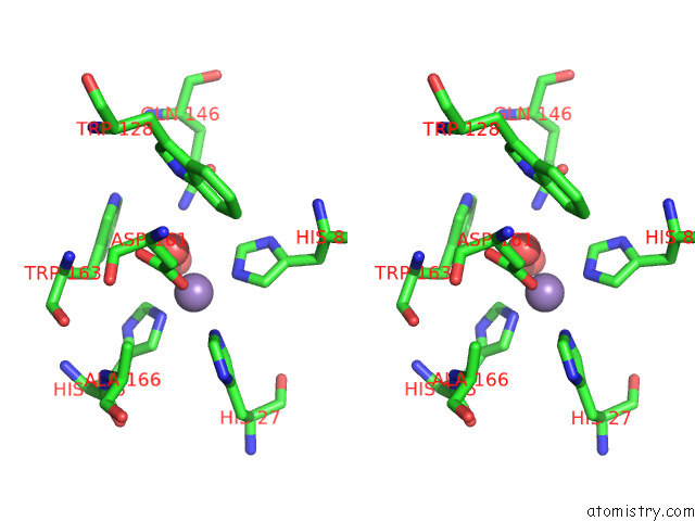
Stereo pair view
Mono view
Stereo pair view
A full contact list of Manganese with other atoms in the Mn binding
site number 4 of Crystal Structure of A Mutant Staphylococcus Equorum Manganese Superoxide Dismutase K38R and A121E within 5.0Å range:
|
Reference:
D.S.Retnoningrum,
H.Yoshida,
A.A.Artarini,
W.T.Ismaya.
Crystal Structure of A Mutant Staphylococcus Equorum Manganese Superoxide Dismutase K38R and A121E To Be Published.
Page generated: Sat Aug 16 23:56:29 2025
Last articles
Na in 6C9NNa in 6C89
Na in 6C96
Na in 6C6F
Na in 6C86
Na in 6C7X
Na in 6C7W
Na in 6C67
Na in 6C5B
Na in 6C6E