Manganese »
PDB 7uuh-7x9i »
7wg0 »
Manganese in PDB 7wg0: Structure of the Manganese Protoporphyrin IX-Reconstituted CYP102A1 Heme Domain with N-Palmitoyl-L-Phenylalanine
Enzymatic activity of Structure of the Manganese Protoporphyrin IX-Reconstituted CYP102A1 Heme Domain with N-Palmitoyl-L-Phenylalanine
All present enzymatic activity of Structure of the Manganese Protoporphyrin IX-Reconstituted CYP102A1 Heme Domain with N-Palmitoyl-L-Phenylalanine:
1.14.14.1; 1.6.2.4;
1.14.14.1; 1.6.2.4;
Protein crystallography data
The structure of Structure of the Manganese Protoporphyrin IX-Reconstituted CYP102A1 Heme Domain with N-Palmitoyl-L-Phenylalanine, PDB code: 7wg0
was solved by
O.Keita,
S.Osami,
A.Yuichiro,
S.Hiroshi,
with X-Ray Crystallography technique. A brief refinement statistics is given in the table below:
| Resolution Low / High (Å) | 48.52 / 2.20 |
| Space group | P 21 21 21 |
| Cell size a, b, c (Å), α, β, γ (°) | 58.78, 127.89, 148.67, 90, 90, 90 |
| R / Rfree (%) | 20.8 / 26.7 |
Manganese Binding Sites:
The binding sites of Manganese atom in the Structure of the Manganese Protoporphyrin IX-Reconstituted CYP102A1 Heme Domain with N-Palmitoyl-L-Phenylalanine
(pdb code 7wg0). This binding sites where shown within
5.0 Angstroms radius around Manganese atom.
In total 2 binding sites of Manganese where determined in the Structure of the Manganese Protoporphyrin IX-Reconstituted CYP102A1 Heme Domain with N-Palmitoyl-L-Phenylalanine, PDB code: 7wg0:
Jump to Manganese binding site number: 1; 2;
In total 2 binding sites of Manganese where determined in the Structure of the Manganese Protoporphyrin IX-Reconstituted CYP102A1 Heme Domain with N-Palmitoyl-L-Phenylalanine, PDB code: 7wg0:
Jump to Manganese binding site number: 1; 2;
Manganese binding site 1 out of 2 in 7wg0
Go back to
Manganese binding site 1 out
of 2 in the Structure of the Manganese Protoporphyrin IX-Reconstituted CYP102A1 Heme Domain with N-Palmitoyl-L-Phenylalanine
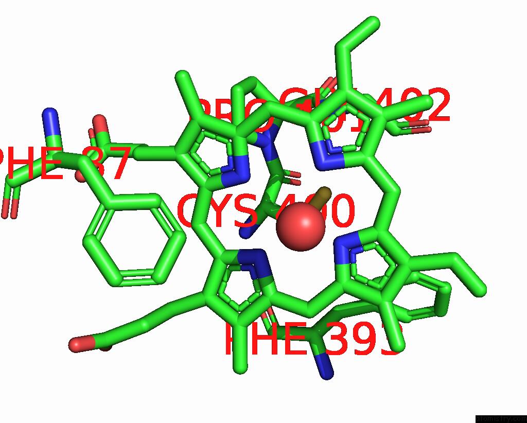
Mono view
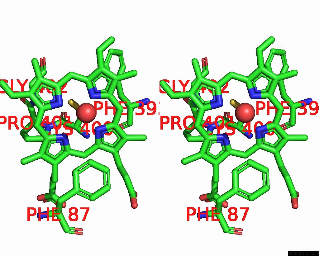
Stereo pair view
Mono view
Stereo pair view
A full contact list of Manganese with other atoms in the Mn binding
site number 1 of Structure of the Manganese Protoporphyrin IX-Reconstituted CYP102A1 Heme Domain with N-Palmitoyl-L-Phenylalanine within 5.0Å range:
|
Manganese binding site 2 out of 2 in 7wg0
Go back to
Manganese binding site 2 out
of 2 in the Structure of the Manganese Protoporphyrin IX-Reconstituted CYP102A1 Heme Domain with N-Palmitoyl-L-Phenylalanine
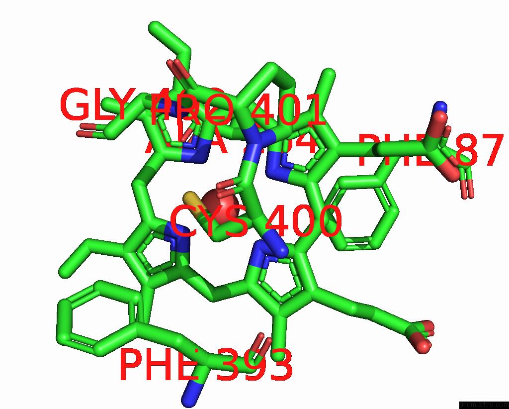
Mono view
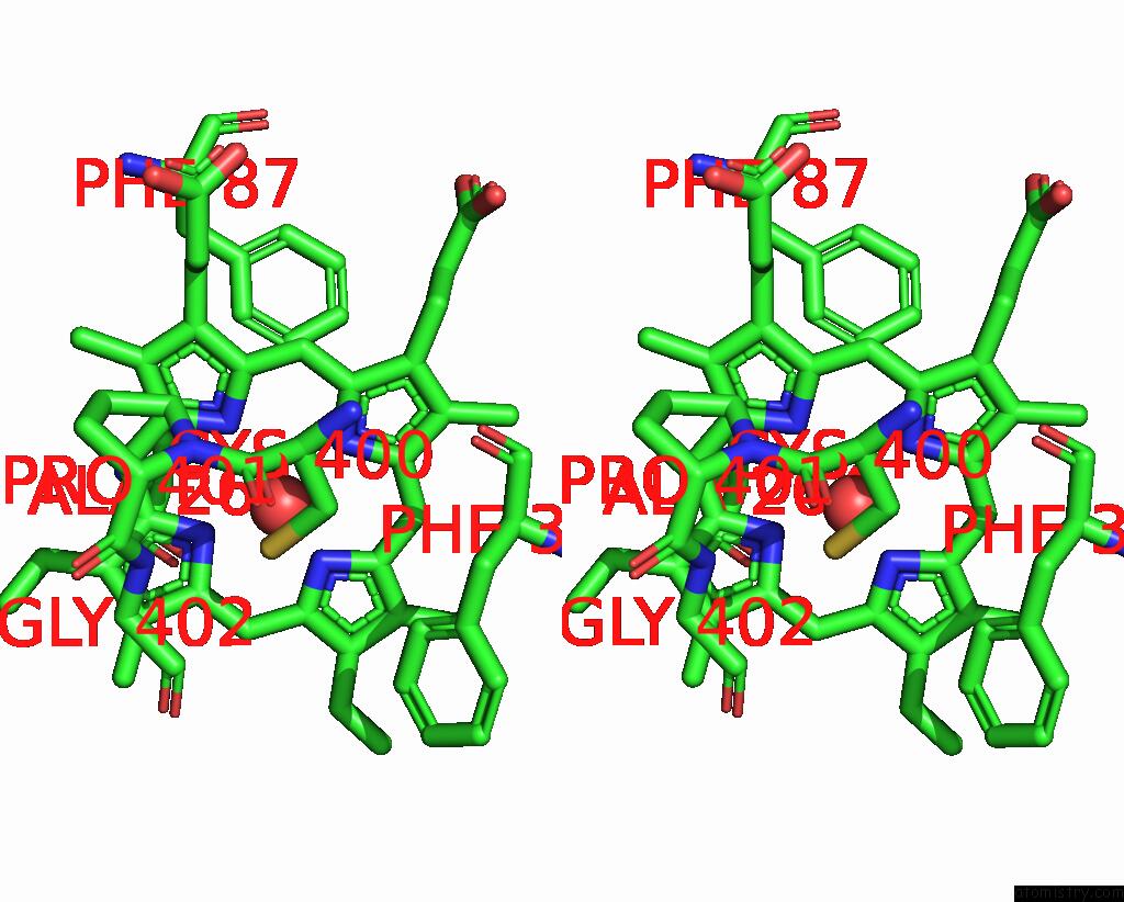
Stereo pair view
Mono view
Stereo pair view
A full contact list of Manganese with other atoms in the Mn binding
site number 2 of Structure of the Manganese Protoporphyrin IX-Reconstituted CYP102A1 Heme Domain with N-Palmitoyl-L-Phenylalanine within 5.0Å range:
|
Reference:
O.Keita,
S.Osami,
A.Yuichiro,
S.Hiroshi.
Structure of the Manganese Protoporphyrin IX-Reconstituted CYP102A1 Heme Domain with N-Palmitoyl-L-Phenylalanine To Be Published.
Page generated: Sat Aug 16 23:55:49 2025
Last articles
Ni in 5J4RNi in 5I8T
Ni in 5I93
Ni in 5I91
Ni in 5I8S
Ni in 5HZU
Ni in 5I14
Ni in 5I0C
Ni in 5HUQ
Ni in 5HT9