Manganese »
PDB 7ohl-7sck »
7omb »
Manganese in PDB 7omb: Crystal Structure of Kod Dna Polymerase in A Ternary Complex with A P/T Duplex Containing An Extended 5' Single Stranded Template Overhang
Enzymatic activity of Crystal Structure of Kod Dna Polymerase in A Ternary Complex with A P/T Duplex Containing An Extended 5' Single Stranded Template Overhang
All present enzymatic activity of Crystal Structure of Kod Dna Polymerase in A Ternary Complex with A P/T Duplex Containing An Extended 5' Single Stranded Template Overhang:
2.7.7.7;
2.7.7.7;
Protein crystallography data
The structure of Crystal Structure of Kod Dna Polymerase in A Ternary Complex with A P/T Duplex Containing An Extended 5' Single Stranded Template Overhang, PDB code: 7omb
was solved by
K.Betz,
H.M.Kropp,
K.Diederichs,
A.Marx,
with X-Ray Crystallography technique. A brief refinement statistics is given in the table below:
| Resolution Low / High (Å) | 46.28 / 2.01 |
| Space group | P 21 21 2 |
| Cell size a, b, c (Å), α, β, γ (°) | 108.081, 147.153, 71.332, 90, 90, 90 |
| R / Rfree (%) | 20 / 23.6 |
Other elements in 7omb:
The structure of Crystal Structure of Kod Dna Polymerase in A Ternary Complex with A P/T Duplex Containing An Extended 5' Single Stranded Template Overhang also contains other interesting chemical elements:
| Magnesium | (Mg) | 2 atoms |
Manganese Binding Sites:
The binding sites of Manganese atom in the Crystal Structure of Kod Dna Polymerase in A Ternary Complex with A P/T Duplex Containing An Extended 5' Single Stranded Template Overhang
(pdb code 7omb). This binding sites where shown within
5.0 Angstroms radius around Manganese atom.
In total 4 binding sites of Manganese where determined in the Crystal Structure of Kod Dna Polymerase in A Ternary Complex with A P/T Duplex Containing An Extended 5' Single Stranded Template Overhang, PDB code: 7omb:
Jump to Manganese binding site number: 1; 2; 3; 4;
In total 4 binding sites of Manganese where determined in the Crystal Structure of Kod Dna Polymerase in A Ternary Complex with A P/T Duplex Containing An Extended 5' Single Stranded Template Overhang, PDB code: 7omb:
Jump to Manganese binding site number: 1; 2; 3; 4;
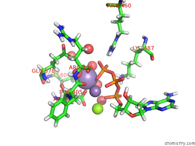
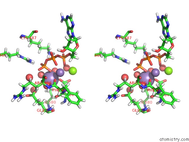
Manganese binding site 1 out of 4 in 7omb
Go back to
Manganese binding site 1 out
of 4 in the Crystal Structure of Kod Dna Polymerase in A Ternary Complex with A P/T Duplex Containing An Extended 5' Single Stranded Template Overhang
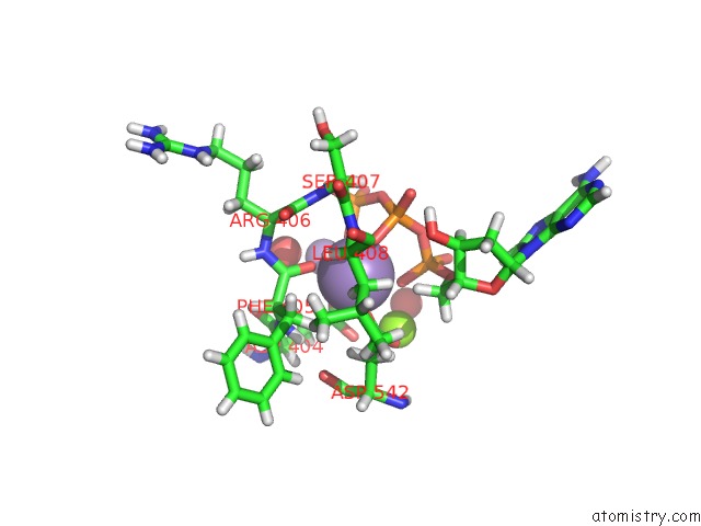
Mono view
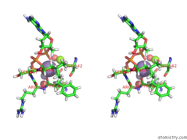
Stereo pair view
Mono view
Stereo pair view
A full contact list of Manganese with other atoms in the Mn binding
site number 1 of Crystal Structure of Kod Dna Polymerase in A Ternary Complex with A P/T Duplex Containing An Extended 5' Single Stranded Template Overhang within 5.0Å range:
|
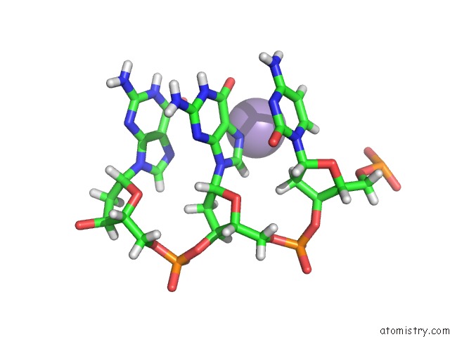
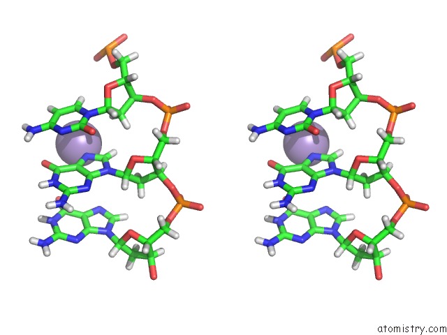
Manganese binding site 2 out of 4 in 7omb
Go back to
Manganese binding site 2 out
of 4 in the Crystal Structure of Kod Dna Polymerase in A Ternary Complex with A P/T Duplex Containing An Extended 5' Single Stranded Template Overhang
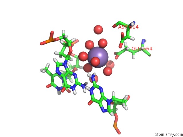
Mono view
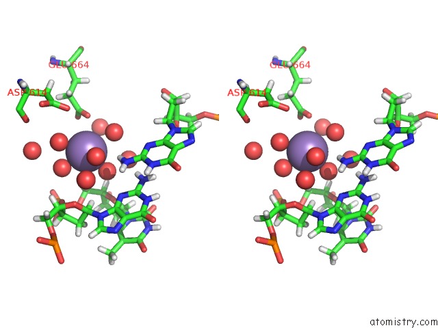
Stereo pair view
Mono view
Stereo pair view
A full contact list of Manganese with other atoms in the Mn binding
site number 2 of Crystal Structure of Kod Dna Polymerase in A Ternary Complex with A P/T Duplex Containing An Extended 5' Single Stranded Template Overhang within 5.0Å range:
|
Manganese binding site 3 out of 4 in 7omb
Go back to
Manganese binding site 3 out
of 4 in the Crystal Structure of Kod Dna Polymerase in A Ternary Complex with A P/T Duplex Containing An Extended 5' Single Stranded Template Overhang
Mono view
Stereo pair view
Mono view
Stereo pair view
A full contact list of Manganese with other atoms in the Mn binding
site number 3 of Crystal Structure of Kod Dna Polymerase in A Ternary Complex with A P/T Duplex Containing An Extended 5' Single Stranded Template Overhang within 5.0Å range:
|
Manganese binding site 4 out of 4 in 7omb
Go back to
Manganese binding site 4 out
of 4 in the Crystal Structure of Kod Dna Polymerase in A Ternary Complex with A P/T Duplex Containing An Extended 5' Single Stranded Template Overhang
Mono view
Stereo pair view
Mono view
Stereo pair view
A full contact list of Manganese with other atoms in the Mn binding
site number 4 of Crystal Structure of Kod Dna Polymerase in A Ternary Complex with A P/T Duplex Containing An Extended 5' Single Stranded Template Overhang within 5.0Å range:
|
Reference:
H.M.Kropp,
S.Ludmann,
K.Diederichs,
K.Betz,
A.Marx.
Structural Basis For the Recognition of Deaminated Nucleobases By An Archaeal Dna Polymerase. Chembiochem 2021.
ISSN: ESSN 1439-7633
PubMed: 34486208
DOI: 10.1002/CBIC.202100306
Page generated: Sun Oct 6 10:22:58 2024
ISSN: ESSN 1439-7633
PubMed: 34486208
DOI: 10.1002/CBIC.202100306
Last articles
K in 2FRZK in 2FPQ
K in 2FPM
K in 2FPL
K in 2FPK
K in 2FPG
K in 2FP4
K in 2FEU
K in 2FFY
K in 2FDE