Manganese »
PDB 7mxs-7ohg »
7n7g »
Manganese in PDB 7n7g: Crystal Structure of Fosb From Enterococcus Faecium with Fosfomycin
Protein crystallography data
The structure of Crystal Structure of Fosb From Enterococcus Faecium with Fosfomycin, PDB code: 7n7g
was solved by
M.R.Shay,
Z.Simmons,
M.K.Thompson,
with X-Ray Crystallography technique. A brief refinement statistics is given in the table below:
| Resolution Low / High (Å) | 23.80 / 2.00 |
| Space group | P 41 21 2 |
| Cell size a, b, c (Å), α, β, γ (°) | 79.874, 79.874, 95.639, 90, 90, 90 |
| R / Rfree (%) | 21.4 / 25.4 |
Manganese Binding Sites:
The binding sites of Manganese atom in the Crystal Structure of Fosb From Enterococcus Faecium with Fosfomycin
(pdb code 7n7g). This binding sites where shown within
5.0 Angstroms radius around Manganese atom.
In total 2 binding sites of Manganese where determined in the Crystal Structure of Fosb From Enterococcus Faecium with Fosfomycin, PDB code: 7n7g:
Jump to Manganese binding site number: 1; 2;
In total 2 binding sites of Manganese where determined in the Crystal Structure of Fosb From Enterococcus Faecium with Fosfomycin, PDB code: 7n7g:
Jump to Manganese binding site number: 1; 2;
Manganese binding site 1 out of 2 in 7n7g
Go back to
Manganese binding site 1 out
of 2 in the Crystal Structure of Fosb From Enterococcus Faecium with Fosfomycin
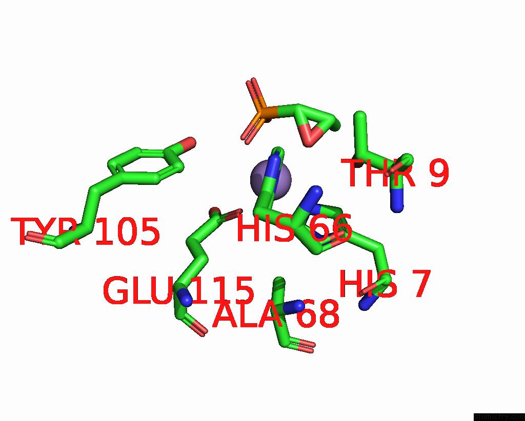
Mono view

Stereo pair view
Mono view
Stereo pair view
A full contact list of Manganese with other atoms in the Mn binding
site number 1 of Crystal Structure of Fosb From Enterococcus Faecium with Fosfomycin within 5.0Å range:
|
Manganese binding site 2 out of 2 in 7n7g
Go back to
Manganese binding site 2 out
of 2 in the Crystal Structure of Fosb From Enterococcus Faecium with Fosfomycin
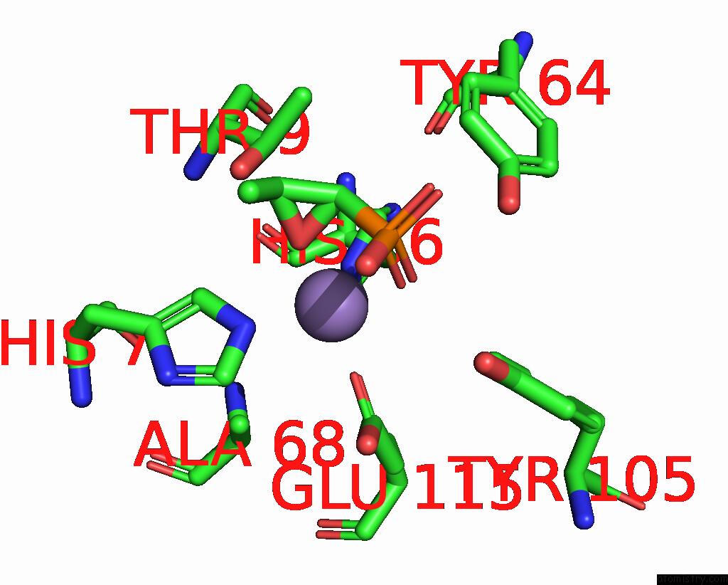
Mono view
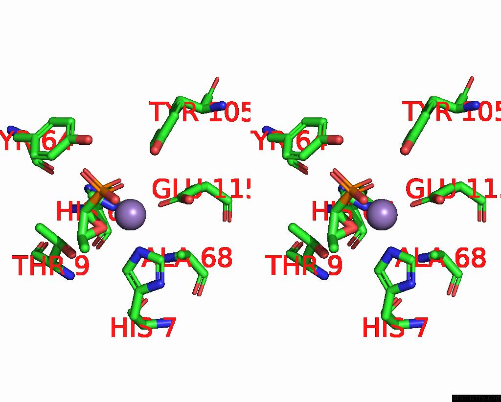
Stereo pair view
Mono view
Stereo pair view
A full contact list of Manganese with other atoms in the Mn binding
site number 2 of Crystal Structure of Fosb From Enterococcus Faecium with Fosfomycin within 5.0Å range:
|
Reference:
V.Wiltsie,
S.Travis,
M.R.Shay,
Z.Simmons,
P.Frantom,
M.K.Thompson.
Structural and Functional Characterization of Fosfomycin Resistance Conferred By Fosb From Enterococcus Faecium. Protein Sci. V. 31 580 2022.
ISSN: ESSN 1469-896X
PubMed: 34882867
DOI: 10.1002/PRO.4253
Page generated: Sun Oct 6 10:15:36 2024
ISSN: ESSN 1469-896X
PubMed: 34882867
DOI: 10.1002/PRO.4253
Last articles
Kr in 6ZMAKr in 6SP8
Kr in 6EYE
Kr in 5D51
Kr in 6RGM
Kr in 6IC1
Kr in 5O17
Kr in 5NW6
Kr in 5FSP
Kr in 5FST