Manganese »
PDB 7mxs-7ohg »
7n02 »
Manganese in PDB 7n02: X-Ray Crystallographic Structure Model of Lactococcus Lactis Prolidase Mutant D36S
Enzymatic activity of X-Ray Crystallographic Structure Model of Lactococcus Lactis Prolidase Mutant D36S
All present enzymatic activity of X-Ray Crystallographic Structure Model of Lactococcus Lactis Prolidase Mutant D36S:
3.4.13.9;
3.4.13.9;
Protein crystallography data
The structure of X-Ray Crystallographic Structure Model of Lactococcus Lactis Prolidase Mutant D36S, PDB code: 7n02
was solved by
S.Xu,
P.Grochulski,
T.Tanaka,
with X-Ray Crystallography technique. A brief refinement statistics is given in the table below:
| Resolution Low / High (Å) | 43.64 / 2.35 |
| Space group | P 21 21 21 |
| Cell size a, b, c (Å), α, β, γ (°) | 83.12, 87.27, 119.4, 90, 90, 90 |
| R / Rfree (%) | 21.2 / 27 |
Manganese Binding Sites:
The binding sites of Manganese atom in the X-Ray Crystallographic Structure Model of Lactococcus Lactis Prolidase Mutant D36S
(pdb code 7n02). This binding sites where shown within
5.0 Angstroms radius around Manganese atom.
In total 4 binding sites of Manganese where determined in the X-Ray Crystallographic Structure Model of Lactococcus Lactis Prolidase Mutant D36S, PDB code: 7n02:
Jump to Manganese binding site number: 1; 2; 3; 4;
In total 4 binding sites of Manganese where determined in the X-Ray Crystallographic Structure Model of Lactococcus Lactis Prolidase Mutant D36S, PDB code: 7n02:
Jump to Manganese binding site number: 1; 2; 3; 4;
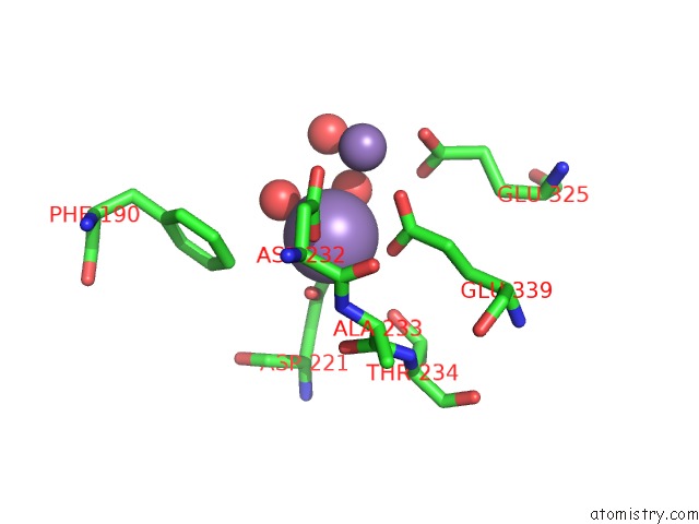
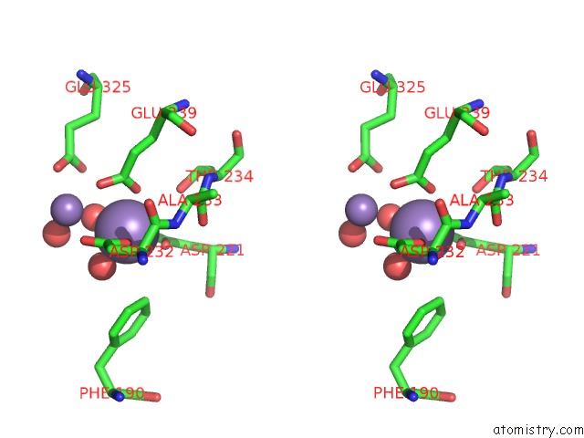
Manganese binding site 1 out of 4 in 7n02
Go back to
Manganese binding site 1 out
of 4 in the X-Ray Crystallographic Structure Model of Lactococcus Lactis Prolidase Mutant D36S
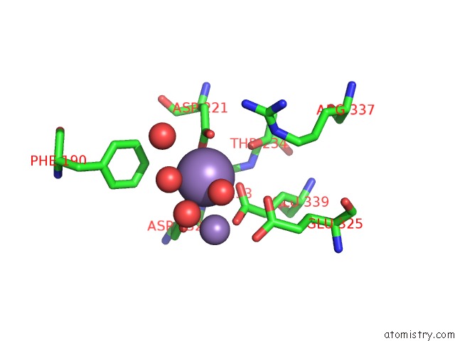
Mono view
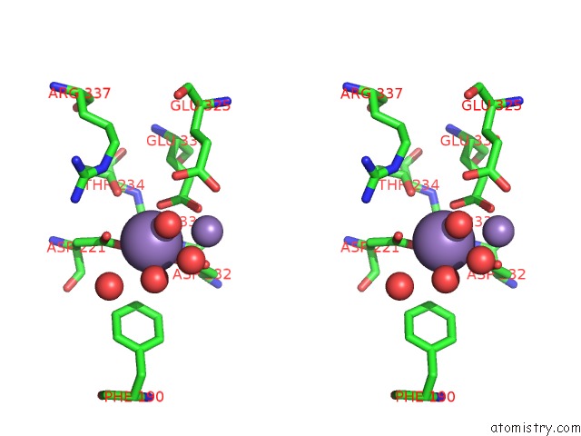
Stereo pair view
Mono view
Stereo pair view
A full contact list of Manganese with other atoms in the Mn binding
site number 1 of X-Ray Crystallographic Structure Model of Lactococcus Lactis Prolidase Mutant D36S within 5.0Å range:
|
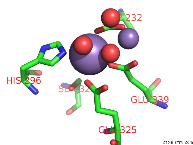
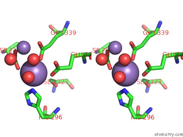
Manganese binding site 2 out of 4 in 7n02
Go back to
Manganese binding site 2 out
of 4 in the X-Ray Crystallographic Structure Model of Lactococcus Lactis Prolidase Mutant D36S
Mono view
Stereo pair view
Mono view
Stereo pair view
A full contact list of Manganese with other atoms in the Mn binding
site number 2 of X-Ray Crystallographic Structure Model of Lactococcus Lactis Prolidase Mutant D36S within 5.0Å range:
|
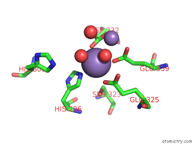
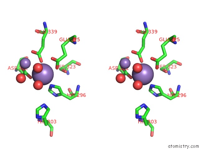
Manganese binding site 3 out of 4 in 7n02
Go back to
Manganese binding site 3 out
of 4 in the X-Ray Crystallographic Structure Model of Lactococcus Lactis Prolidase Mutant D36S
Mono view
Stereo pair view
Mono view
Stereo pair view
A full contact list of Manganese with other atoms in the Mn binding
site number 3 of X-Ray Crystallographic Structure Model of Lactococcus Lactis Prolidase Mutant D36S within 5.0Å range:
|
Manganese binding site 4 out of 4 in 7n02
Go back to
Manganese binding site 4 out
of 4 in the X-Ray Crystallographic Structure Model of Lactococcus Lactis Prolidase Mutant D36S
Mono view
Stereo pair view
Mono view
Stereo pair view
A full contact list of Manganese with other atoms in the Mn binding
site number 4 of X-Ray Crystallographic Structure Model of Lactococcus Lactis Prolidase Mutant D36S within 5.0Å range:
|
Reference:
O.Kgosisejo,
J.A.Chen,
P.Grochulski,
T.Tanaka.
Crystallographic Structure of Recombinant Lactococcus Lactis Prolidase to Support Proposed Structure-Function Relationships. Biochim Biophys Acta V.1865 473 2017PROTEINS Proteom.
ISSN: ISSN 1570-9639
PubMed: 28179139
DOI: 10.1016/J.BBAPAP.2017.02.004
Page generated: Sun Oct 6 10:13:10 2024
ISSN: ISSN 1570-9639
PubMed: 28179139
DOI: 10.1016/J.BBAPAP.2017.02.004
Last articles
K in 7QQQK in 7QQP
K in 7QQO
K in 7QK5
K in 7QIX
K in 7QNO
K in 7QIY
K in 7Q3X
K in 7QDN
K in 7QF6