Manganese »
PDB 7ksu-7lrd »
7lol »
Manganese in PDB 7lol: The Structure of Agmatinase From E. Coli at 1.8 A Displaying Urea and Agmatine
Enzymatic activity of The Structure of Agmatinase From E. Coli at 1.8 A Displaying Urea and Agmatine
All present enzymatic activity of The Structure of Agmatinase From E. Coli at 1.8 A Displaying Urea and Agmatine:
3.5.3.11;
3.5.3.11;
Protein crystallography data
The structure of The Structure of Agmatinase From E. Coli at 1.8 A Displaying Urea and Agmatine, PDB code: 7lol
was solved by
P.Maturana,
M.Figueroa,
F.Gonzalez-Ordenes,
P.Villalobos,
J.Martinez-Oyanedel,
E.A.Uribe,
V.Castro-Fernandez,
with X-Ray Crystallography technique. A brief refinement statistics is given in the table below:
| Resolution Low / High (Å) | 35.79 / 1.80 |
| Space group | H 3 2 |
| Cell size a, b, c (Å), α, β, γ (°) | 81.746, 81.746, 207.436, 90, 90, 120 |
| R / Rfree (%) | 17 / 19.5 |
Manganese Binding Sites:
The binding sites of Manganese atom in the The Structure of Agmatinase From E. Coli at 1.8 A Displaying Urea and Agmatine
(pdb code 7lol). This binding sites where shown within
5.0 Angstroms radius around Manganese atom.
In total 3 binding sites of Manganese where determined in the The Structure of Agmatinase From E. Coli at 1.8 A Displaying Urea and Agmatine, PDB code: 7lol:
Jump to Manganese binding site number: 1; 2; 3;
In total 3 binding sites of Manganese where determined in the The Structure of Agmatinase From E. Coli at 1.8 A Displaying Urea and Agmatine, PDB code: 7lol:
Jump to Manganese binding site number: 1; 2; 3;
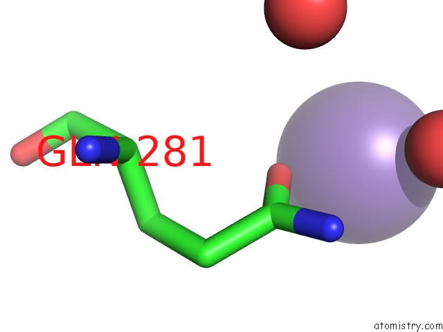
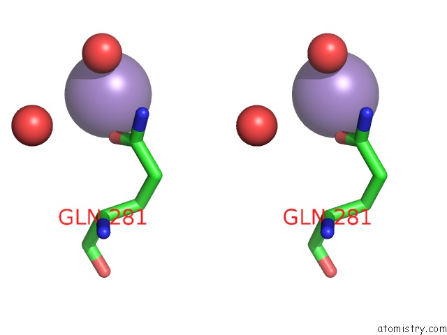
Manganese binding site 1 out of 3 in 7lol
Go back to
Manganese binding site 1 out
of 3 in the The Structure of Agmatinase From E. Coli at 1.8 A Displaying Urea and Agmatine
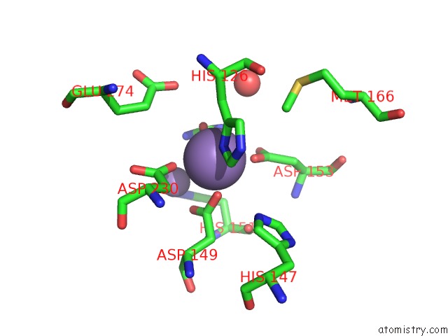
Mono view
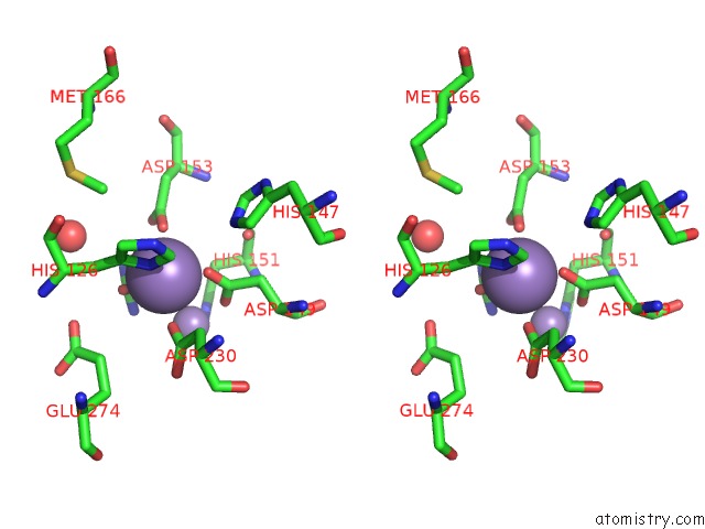
Stereo pair view
Mono view
Stereo pair view
A full contact list of Manganese with other atoms in the Mn binding
site number 1 of The Structure of Agmatinase From E. Coli at 1.8 A Displaying Urea and Agmatine within 5.0Å range:
|
Manganese binding site 2 out of 3 in 7lol
Go back to
Manganese binding site 2 out
of 3 in the The Structure of Agmatinase From E. Coli at 1.8 A Displaying Urea and Agmatine
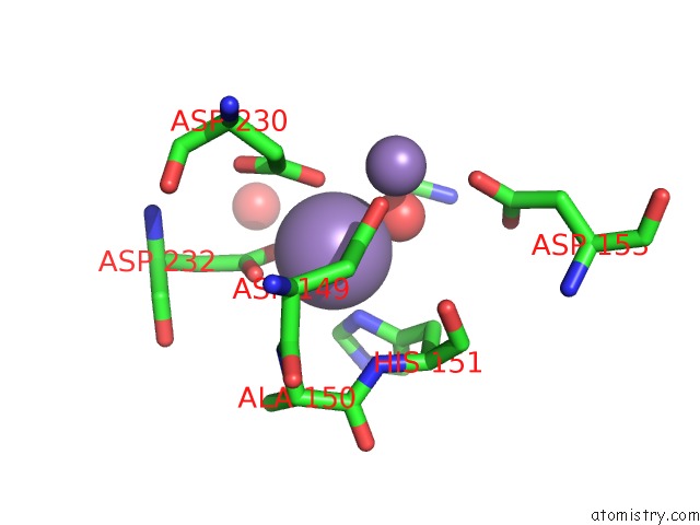
Mono view
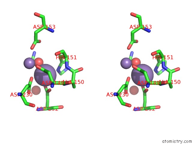
Stereo pair view
Mono view
Stereo pair view
A full contact list of Manganese with other atoms in the Mn binding
site number 2 of The Structure of Agmatinase From E. Coli at 1.8 A Displaying Urea and Agmatine within 5.0Å range:
|
Manganese binding site 3 out of 3 in 7lol
Go back to
Manganese binding site 3 out
of 3 in the The Structure of Agmatinase From E. Coli at 1.8 A Displaying Urea and Agmatine
Mono view
Stereo pair view
Mono view
Stereo pair view
A full contact list of Manganese with other atoms in the Mn binding
site number 3 of The Structure of Agmatinase From E. Coli at 1.8 A Displaying Urea and Agmatine within 5.0Å range:
|
Reference:
P.Maturana,
M.S.Orellana,
S.M.Herrera,
I.Martinez,
M.Figueroa,
J.Martinez-Oyanedel,
V.Castro-Fernandez,
E.Uribe.
Crystal Structure of Escherichia Coli Agmatinase: Catalytic Mechanism and Residues Relevant For Substrate Specificity Int J Mol Sci V. 22 2021.
ISSN: ESSN 1422-0067
DOI: 10.3390/IJMS22094769
Page generated: Sun Oct 6 09:54:24 2024
ISSN: ESSN 1422-0067
DOI: 10.3390/IJMS22094769
Last articles
Mg in 2D0QMg in 2D2V
Mg in 2D0O
Mg in 2D2F
Mg in 2D2C
Mg in 2D25
Mg in 2D1K
Mg in 2CW6
Mg in 2D0B
Mg in 2CZ6