Manganese »
PDB 6zbn-7bm0 »
7a13 »
Manganese in PDB 7a13: Crystal Structure of Human Methionine Aminopeptidase-2 in Complex with An Inhibitor GSK1978537A (Compound 27)
Enzymatic activity of Crystal Structure of Human Methionine Aminopeptidase-2 in Complex with An Inhibitor GSK1978537A (Compound 27)
All present enzymatic activity of Crystal Structure of Human Methionine Aminopeptidase-2 in Complex with An Inhibitor GSK1978537A (Compound 27):
3.4.11.18;
3.4.11.18;
Protein crystallography data
The structure of Crystal Structure of Human Methionine Aminopeptidase-2 in Complex with An Inhibitor GSK1978537A (Compound 27), PDB code: 7a13
was solved by
J.H.Thorpe,
with X-Ray Crystallography technique. A brief refinement statistics is given in the table below:
| Resolution Low / High (Å) | 29.97 / 2.04 |
| Space group | C 2 2 21 |
| Cell size a, b, c (Å), α, β, γ (°) | 89.195, 100.770, 99.396, 90.00, 90.00, 90.00 |
| R / Rfree (%) | 16.6 / 19.7 |
Other elements in 7a13:
The structure of Crystal Structure of Human Methionine Aminopeptidase-2 in Complex with An Inhibitor GSK1978537A (Compound 27) also contains other interesting chemical elements:
| Chlorine | (Cl) | 1 atom |
Manganese Binding Sites:
The binding sites of Manganese atom in the Crystal Structure of Human Methionine Aminopeptidase-2 in Complex with An Inhibitor GSK1978537A (Compound 27)
(pdb code 7a13). This binding sites where shown within
5.0 Angstroms radius around Manganese atom.
In total 2 binding sites of Manganese where determined in the Crystal Structure of Human Methionine Aminopeptidase-2 in Complex with An Inhibitor GSK1978537A (Compound 27), PDB code: 7a13:
Jump to Manganese binding site number: 1; 2;
In total 2 binding sites of Manganese where determined in the Crystal Structure of Human Methionine Aminopeptidase-2 in Complex with An Inhibitor GSK1978537A (Compound 27), PDB code: 7a13:
Jump to Manganese binding site number: 1; 2;
Manganese binding site 1 out of 2 in 7a13
Go back to
Manganese binding site 1 out
of 2 in the Crystal Structure of Human Methionine Aminopeptidase-2 in Complex with An Inhibitor GSK1978537A (Compound 27)
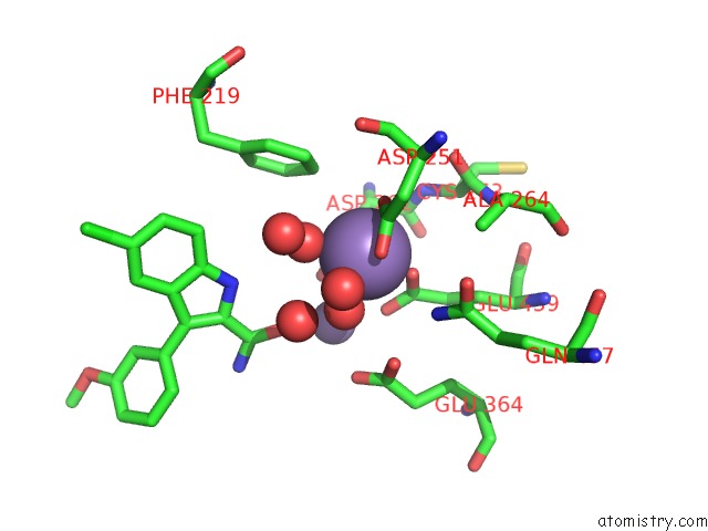
Mono view
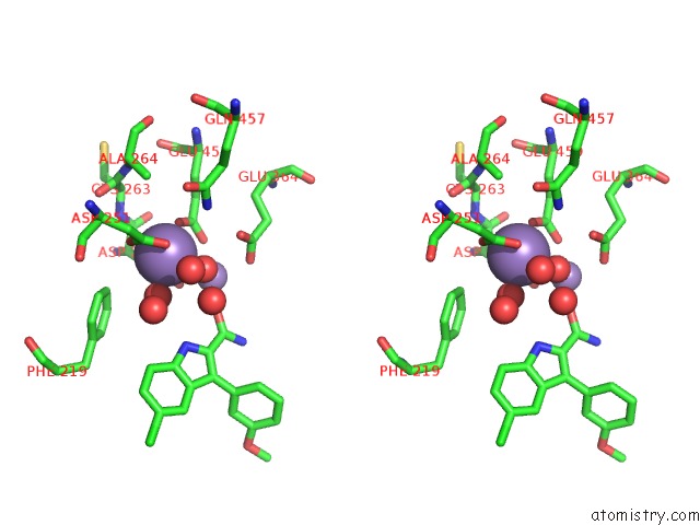
Stereo pair view
Mono view
Stereo pair view
A full contact list of Manganese with other atoms in the Mn binding
site number 1 of Crystal Structure of Human Methionine Aminopeptidase-2 in Complex with An Inhibitor GSK1978537A (Compound 27) within 5.0Å range:
|
Manganese binding site 2 out of 2 in 7a13
Go back to
Manganese binding site 2 out
of 2 in the Crystal Structure of Human Methionine Aminopeptidase-2 in Complex with An Inhibitor GSK1978537A (Compound 27)
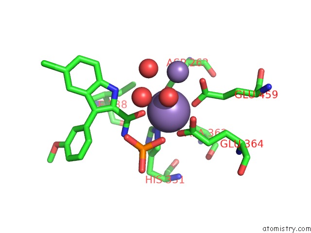
Mono view
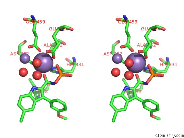
Stereo pair view
Mono view
Stereo pair view
A full contact list of Manganese with other atoms in the Mn binding
site number 2 of Crystal Structure of Human Methionine Aminopeptidase-2 in Complex with An Inhibitor GSK1978537A (Compound 27) within 5.0Å range:
|
Reference:
D.J.Hirst,
M.Brandt,
G.Bruton,
E.Christodoulou,
L.Cutler,
N.Deeks,
J.D.Goodacre,
T.Jack,
M.Lindon,
A.Miah,
K.Page,
N.Parr,
L.Shukla,
M.Sims,
P.Thomas,
J.Thorpe,
D.S.Holmes.
Structure-Based Optimisation of Orally Active & Reversible Metap-2 Inhibitors Maintaining A Tight 'Molecular Budget'. Bioorg.Med.Chem.Lett. V. 30 27533 2020.
ISSN: ESSN 1464-3405
PubMed: 32919012
DOI: 10.1016/J.BMCL.2020.127533
Page generated: Sun Oct 6 08:01:15 2024
ISSN: ESSN 1464-3405
PubMed: 32919012
DOI: 10.1016/J.BMCL.2020.127533
Last articles
K in 3MENK in 3MLB
K in 3MIO
K in 3MIJ
K in 3MD7
K in 3M62
K in 3M63
K in 3LNM
K in 3LUT
K in 3LIB