Manganese »
PDB 6wj4-6z6r »
6wn6 »
Manganese in PDB 6wn6: Crystal Structure of 3-Keto-D-Glucoside 4-Epimerase, Ycjr, From E. Coli, Apo Form
Protein crystallography data
The structure of Crystal Structure of 3-Keto-D-Glucoside 4-Epimerase, Ycjr, From E. Coli, Apo Form, PDB code: 6wn6
was solved by
M.F.Mabanglo,
F.M.Raushel,
K.Mukherjee,
with X-Ray Crystallography technique. A brief refinement statistics is given in the table below:
| Resolution Low / High (Å) | 35.91 / 1.86 |
| Space group | P 41 21 2 |
| Cell size a, b, c (Å), α, β, γ (°) | 116.576, 116.576, 247.725, 90.00, 90.00, 90.00 |
| R / Rfree (%) | 17.6 / 19.9 |
Manganese Binding Sites:
The binding sites of Manganese atom in the Crystal Structure of 3-Keto-D-Glucoside 4-Epimerase, Ycjr, From E. Coli, Apo Form
(pdb code 6wn6). This binding sites where shown within
5.0 Angstroms radius around Manganese atom.
In total 4 binding sites of Manganese where determined in the Crystal Structure of 3-Keto-D-Glucoside 4-Epimerase, Ycjr, From E. Coli, Apo Form, PDB code: 6wn6:
Jump to Manganese binding site number: 1; 2; 3; 4;
In total 4 binding sites of Manganese where determined in the Crystal Structure of 3-Keto-D-Glucoside 4-Epimerase, Ycjr, From E. Coli, Apo Form, PDB code: 6wn6:
Jump to Manganese binding site number: 1; 2; 3; 4;
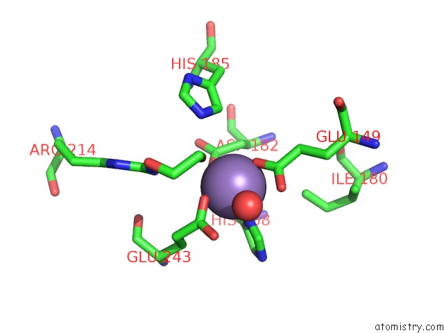
Manganese binding site 1 out of 4 in 6wn6
Go back to
Manganese binding site 1 out
of 4 in the Crystal Structure of 3-Keto-D-Glucoside 4-Epimerase, Ycjr, From E. Coli, Apo Form
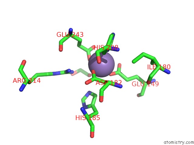
Mono view
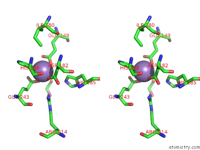
Stereo pair view
Mono view
Stereo pair view
A full contact list of Manganese with other atoms in the Mn binding
site number 1 of Crystal Structure of 3-Keto-D-Glucoside 4-Epimerase, Ycjr, From E. Coli, Apo Form within 5.0Å range:
|
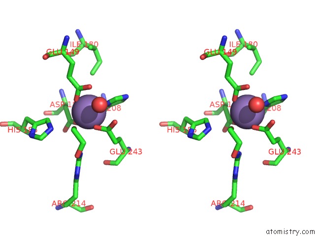
Manganese binding site 2 out of 4 in 6wn6
Go back to
Manganese binding site 2 out
of 4 in the Crystal Structure of 3-Keto-D-Glucoside 4-Epimerase, Ycjr, From E. Coli, Apo Form
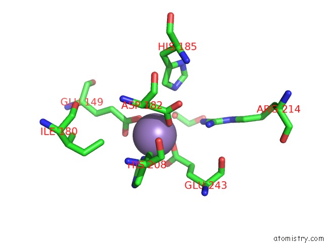
Mono view
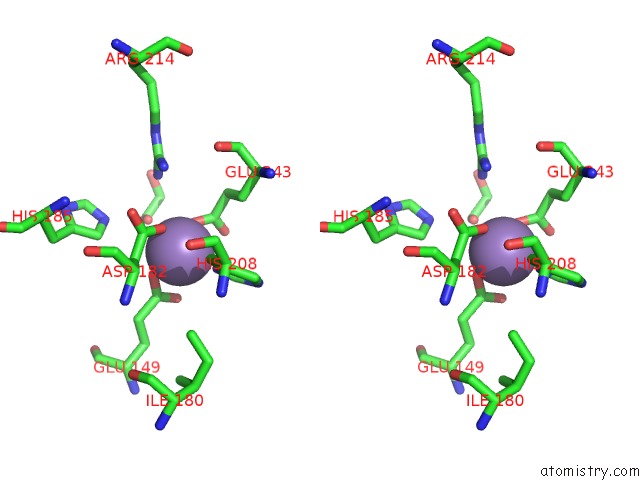
Stereo pair view
Mono view
Stereo pair view
A full contact list of Manganese with other atoms in the Mn binding
site number 2 of Crystal Structure of 3-Keto-D-Glucoside 4-Epimerase, Ycjr, From E. Coli, Apo Form within 5.0Å range:
|
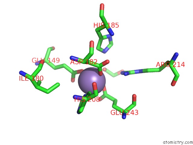
Manganese binding site 3 out of 4 in 6wn6
Go back to
Manganese binding site 3 out
of 4 in the Crystal Structure of 3-Keto-D-Glucoside 4-Epimerase, Ycjr, From E. Coli, Apo Form
Mono view
Stereo pair view
Mono view
Stereo pair view
A full contact list of Manganese with other atoms in the Mn binding
site number 3 of Crystal Structure of 3-Keto-D-Glucoside 4-Epimerase, Ycjr, From E. Coli, Apo Form within 5.0Å range:
|
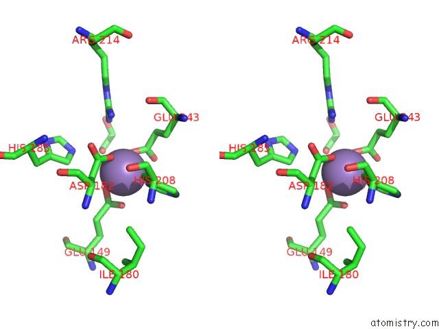
Manganese binding site 4 out of 4 in 6wn6
Go back to
Manganese binding site 4 out
of 4 in the Crystal Structure of 3-Keto-D-Glucoside 4-Epimerase, Ycjr, From E. Coli, Apo Form
Mono view
Stereo pair view
Mono view
Stereo pair view
A full contact list of Manganese with other atoms in the Mn binding
site number 4 of Crystal Structure of 3-Keto-D-Glucoside 4-Epimerase, Ycjr, From E. Coli, Apo Form within 5.0Å range:
|
Reference:
M.F.Mabanglo,
J.P.Huddleston,
K.Mukherjee,
Z.W.Taylor,
F.M.Raushel.
Structure and Reaction Mechanism of Ycjr, An Epimerase That Facilitates the Interconversion of D-Gulosides to D-Glucosides Inescherichia Coli. Biochemistry 2020.
ISSN: ISSN 0006-2960
PubMed: 32437133
DOI: 10.1021/ACS.BIOCHEM.0C00334
Page generated: Sun Oct 6 07:51:04 2024
ISSN: ISSN 0006-2960
PubMed: 32437133
DOI: 10.1021/ACS.BIOCHEM.0C00334
Last articles
K in 6DVNK in 6DVM
K in 6DWO
K in 6DXV
K in 6DVX
K in 6DVL
K in 6DVO
K in 6DUR
K in 6DPN
K in 6DPH