Manganese »
PDB 6vf5-6wij »
6wbt »
Manganese in PDB 6wbt: 2.52 Angstrom Resolution Crystal Structure of 6-Phospho-Alpha- Glucosidase From Gut Microorganisms in Complex with Nad and Glucose- 6-Phosphate
Protein crystallography data
The structure of 2.52 Angstrom Resolution Crystal Structure of 6-Phospho-Alpha- Glucosidase From Gut Microorganisms in Complex with Nad and Glucose- 6-Phosphate, PDB code: 6wbt
was solved by
R.Wu,
Y.Kim,
M.Endres,
J.Joachimiak,
with X-Ray Crystallography technique. A brief refinement statistics is given in the table below:
| Resolution Low / High (Å) | 70.92 / 2.52 |
| Space group | P 21 21 21 |
| Cell size a, b, c (Å), α, β, γ (°) | 102.797, 219.724, 97.978, 90, 90, 90 |
| R / Rfree (%) | 22.7 / 26.6 |
Manganese Binding Sites:
The binding sites of Manganese atom in the 2.52 Angstrom Resolution Crystal Structure of 6-Phospho-Alpha- Glucosidase From Gut Microorganisms in Complex with Nad and Glucose- 6-Phosphate
(pdb code 6wbt). This binding sites where shown within
5.0 Angstroms radius around Manganese atom.
In total 4 binding sites of Manganese where determined in the 2.52 Angstrom Resolution Crystal Structure of 6-Phospho-Alpha- Glucosidase From Gut Microorganisms in Complex with Nad and Glucose- 6-Phosphate, PDB code: 6wbt:
Jump to Manganese binding site number: 1; 2; 3; 4;
In total 4 binding sites of Manganese where determined in the 2.52 Angstrom Resolution Crystal Structure of 6-Phospho-Alpha- Glucosidase From Gut Microorganisms in Complex with Nad and Glucose- 6-Phosphate, PDB code: 6wbt:
Jump to Manganese binding site number: 1; 2; 3; 4;
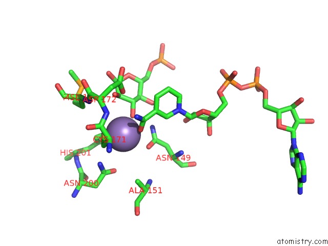
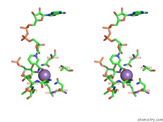
Manganese binding site 1 out of 4 in 6wbt
Go back to
Manganese binding site 1 out
of 4 in the 2.52 Angstrom Resolution Crystal Structure of 6-Phospho-Alpha- Glucosidase From Gut Microorganisms in Complex with Nad and Glucose- 6-Phosphate
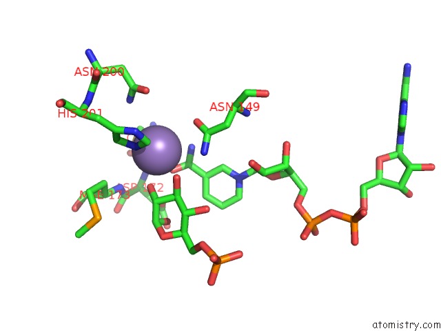
Mono view
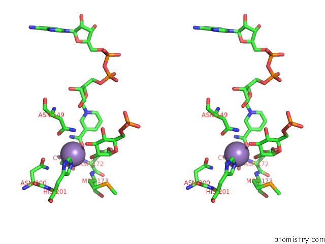
Stereo pair view
Mono view
Stereo pair view
A full contact list of Manganese with other atoms in the Mn binding
site number 1 of 2.52 Angstrom Resolution Crystal Structure of 6-Phospho-Alpha- Glucosidase From Gut Microorganisms in Complex with Nad and Glucose- 6-Phosphate within 5.0Å range:
|
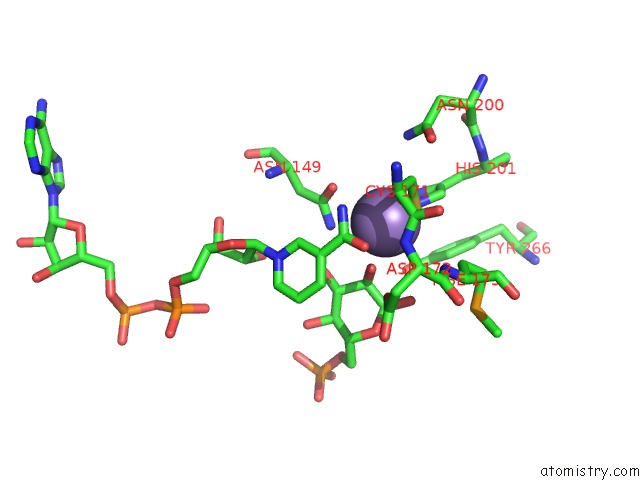
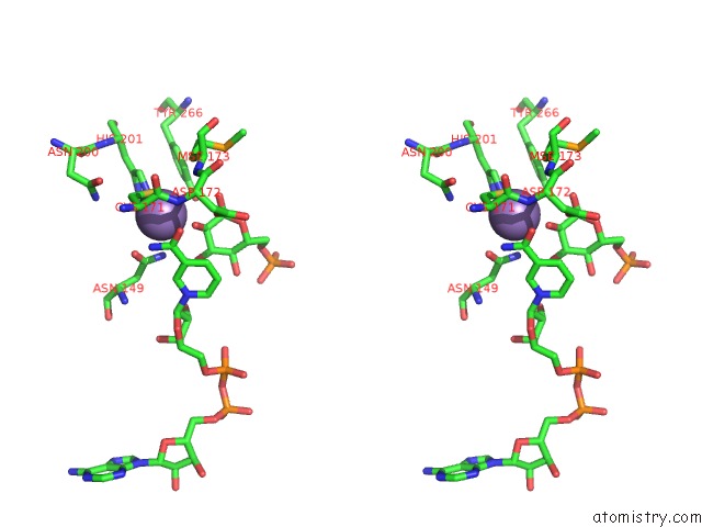
Manganese binding site 2 out of 4 in 6wbt
Go back to
Manganese binding site 2 out
of 4 in the 2.52 Angstrom Resolution Crystal Structure of 6-Phospho-Alpha- Glucosidase From Gut Microorganisms in Complex with Nad and Glucose- 6-Phosphate
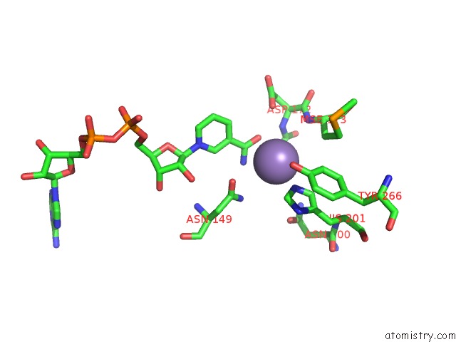
Mono view
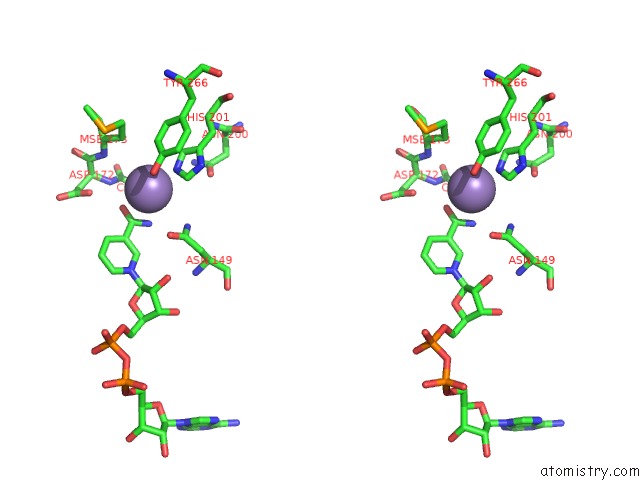
Stereo pair view
Mono view
Stereo pair view
A full contact list of Manganese with other atoms in the Mn binding
site number 2 of 2.52 Angstrom Resolution Crystal Structure of 6-Phospho-Alpha- Glucosidase From Gut Microorganisms in Complex with Nad and Glucose- 6-Phosphate within 5.0Å range:
|
Manganese binding site 3 out of 4 in 6wbt
Go back to
Manganese binding site 3 out
of 4 in the 2.52 Angstrom Resolution Crystal Structure of 6-Phospho-Alpha- Glucosidase From Gut Microorganisms in Complex with Nad and Glucose- 6-Phosphate
Mono view
Stereo pair view
Mono view
Stereo pair view
A full contact list of Manganese with other atoms in the Mn binding
site number 3 of 2.52 Angstrom Resolution Crystal Structure of 6-Phospho-Alpha- Glucosidase From Gut Microorganisms in Complex with Nad and Glucose- 6-Phosphate within 5.0Å range:
|
Manganese binding site 4 out of 4 in 6wbt
Go back to
Manganese binding site 4 out
of 4 in the 2.52 Angstrom Resolution Crystal Structure of 6-Phospho-Alpha- Glucosidase From Gut Microorganisms in Complex with Nad and Glucose- 6-Phosphate
Mono view
Stereo pair view
Mono view
Stereo pair view
A full contact list of Manganese with other atoms in the Mn binding
site number 4 of 2.52 Angstrom Resolution Crystal Structure of 6-Phospho-Alpha- Glucosidase From Gut Microorganisms in Complex with Nad and Glucose- 6-Phosphate within 5.0Å range:
|
Reference:
R.Wu,
Y.Kim,
M.Endres,
J.Joachimiak.
2.52 Angstrom Resolution Crystal Structure of 6-Phospho-Alpha-Glucosidase From Gut Microorganisms in Complex with Nad and Glucose-6-Phosphate To Be Published.
Page generated: Sun Oct 6 07:42:19 2024
Last articles
K in 6R3FK in 6R34
K in 6R33
K in 6R16
K in 6R32
K in 6R30
K in 6R2Z
K in 6R2T
K in 6R2R
K in 6R2P