Manganese »
PDB 6tzp-6vf4 »
6vbz »
Manganese in PDB 6vbz: Crystal Structure of the Rat Mlkl Pseudokinase Domain
Protein crystallography data
The structure of Crystal Structure of the Rat Mlkl Pseudokinase Domain, PDB code: 6vbz
was solved by
K.A.Davies,
P.E.Czabotar,
with X-Ray Crystallography technique. A brief refinement statistics is given in the table below:
| Resolution Low / High (Å) | 48.58 / 2.19 |
| Space group | P 61 2 2 |
| Cell size a, b, c (Å), α, β, γ (°) | 190.057, 190.057, 77.759, 90.00, 90.00, 120.00 |
| R / Rfree (%) | 18.1 / 21.5 |
Manganese Binding Sites:
The binding sites of Manganese atom in the Crystal Structure of the Rat Mlkl Pseudokinase Domain
(pdb code 6vbz). This binding sites where shown within
5.0 Angstroms radius around Manganese atom.
In total 6 binding sites of Manganese where determined in the Crystal Structure of the Rat Mlkl Pseudokinase Domain, PDB code: 6vbz:
Jump to Manganese binding site number: 1; 2; 3; 4; 5; 6;
In total 6 binding sites of Manganese where determined in the Crystal Structure of the Rat Mlkl Pseudokinase Domain, PDB code: 6vbz:
Jump to Manganese binding site number: 1; 2; 3; 4; 5; 6;
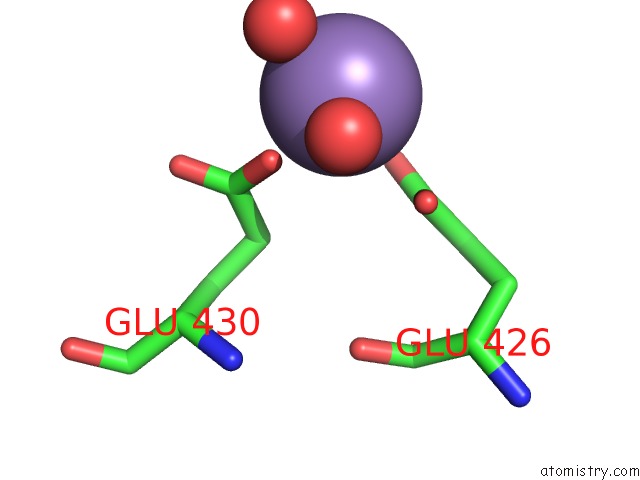
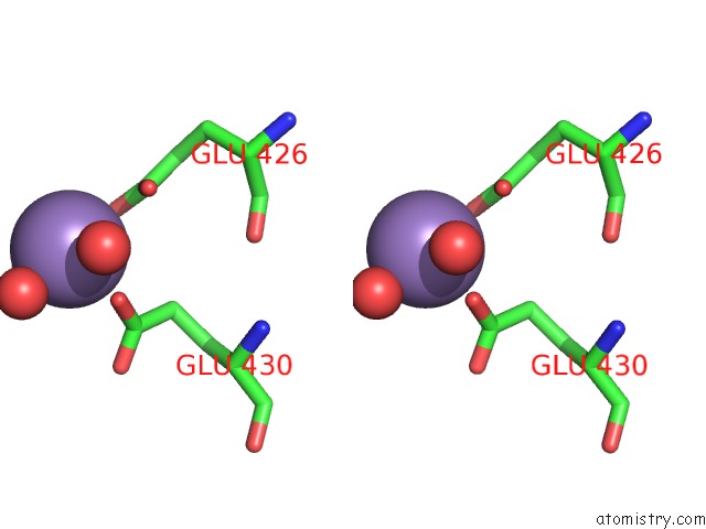
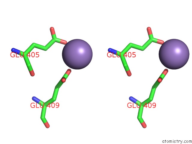
Manganese binding site 1 out of 6 in 6vbz
Go back to
Manganese binding site 1 out
of 6 in the Crystal Structure of the Rat Mlkl Pseudokinase Domain
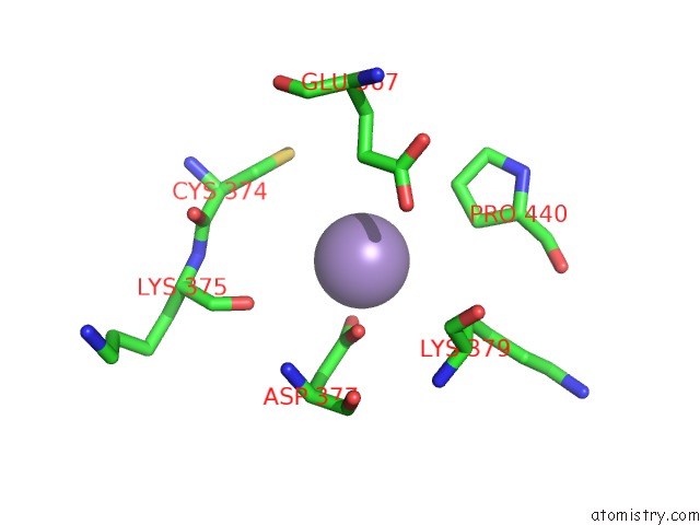
Mono view
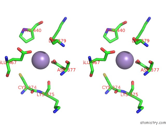
Stereo pair view
Mono view
Stereo pair view
A full contact list of Manganese with other atoms in the Mn binding
site number 1 of Crystal Structure of the Rat Mlkl Pseudokinase Domain within 5.0Å range:
|
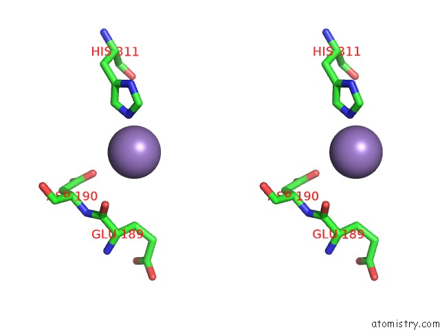
Manganese binding site 2 out of 6 in 6vbz
Go back to
Manganese binding site 2 out
of 6 in the Crystal Structure of the Rat Mlkl Pseudokinase Domain
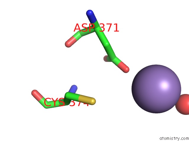
Mono view
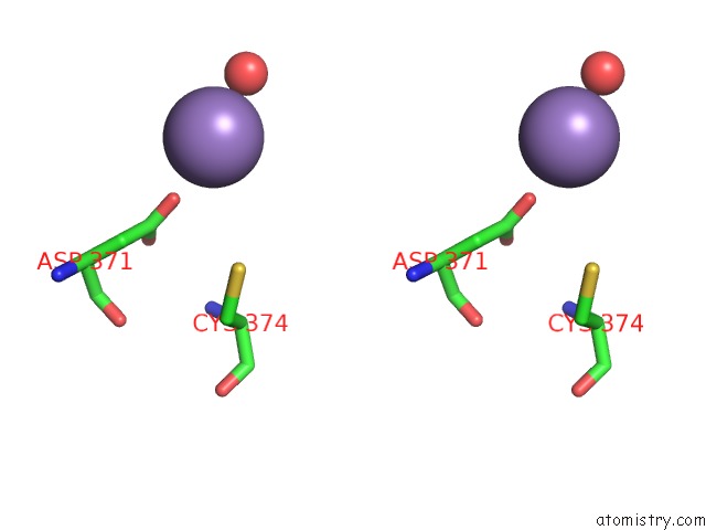
Stereo pair view
Mono view
Stereo pair view
A full contact list of Manganese with other atoms in the Mn binding
site number 2 of Crystal Structure of the Rat Mlkl Pseudokinase Domain within 5.0Å range:
|
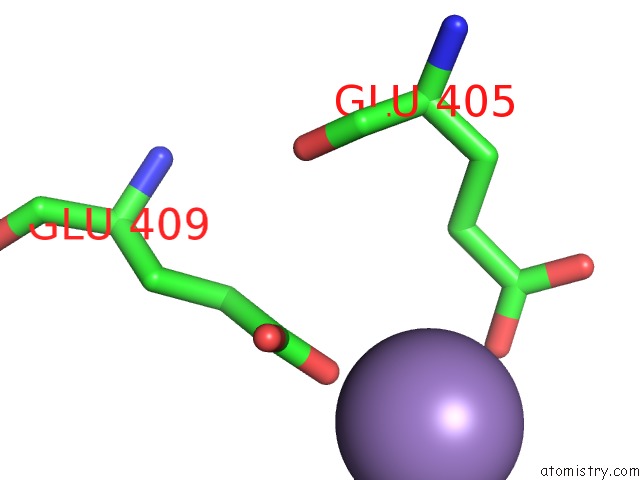
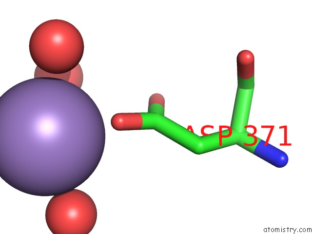
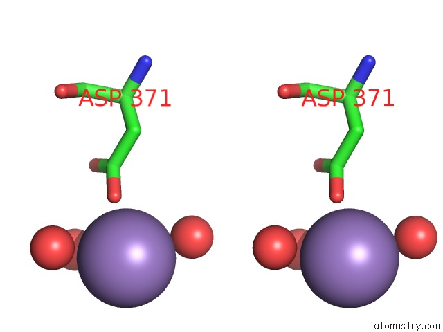
Manganese binding site 3 out of 6 in 6vbz
Go back to
Manganese binding site 3 out
of 6 in the Crystal Structure of the Rat Mlkl Pseudokinase Domain
Mono view
Stereo pair view
Mono view
Stereo pair view
A full contact list of Manganese with other atoms in the Mn binding
site number 3 of Crystal Structure of the Rat Mlkl Pseudokinase Domain within 5.0Å range:
|
Manganese binding site 4 out of 6 in 6vbz
Go back to
Manganese binding site 4 out
of 6 in the Crystal Structure of the Rat Mlkl Pseudokinase Domain

Mono view
Stereo pair view
Mono view
Stereo pair view
A full contact list of Manganese with other atoms in the Mn binding
site number 4 of Crystal Structure of the Rat Mlkl Pseudokinase Domain within 5.0Å range:
|
Manganese binding site 5 out of 6 in 6vbz
Go back to
Manganese binding site 5 out
of 6 in the Crystal Structure of the Rat Mlkl Pseudokinase Domain
Mono view
Stereo pair view
Mono view
Stereo pair view
A full contact list of Manganese with other atoms in the Mn binding
site number 5 of Crystal Structure of the Rat Mlkl Pseudokinase Domain within 5.0Å range:
|
Manganese binding site 6 out of 6 in 6vbz
Go back to
Manganese binding site 6 out
of 6 in the Crystal Structure of the Rat Mlkl Pseudokinase Domain
Mono view
Stereo pair view
Mono view
Stereo pair view
A full contact list of Manganese with other atoms in the Mn binding
site number 6 of Crystal Structure of the Rat Mlkl Pseudokinase Domain within 5.0Å range:
|
Reference:
K.A.Davies,
C.Fitzgibbon,
S.N.Young,
S.E.Garnish,
W.Yeung,
D.Coursier,
R.W.Birkinshaw,
J.J.Sandow,
W.I.L.Lehmann,
L.Y.Liang,
I.S.Lucet,
J.D.Chalmers,
W.M.Patrick,
N.Kannan,
E.J.Petrie,
P.E.Czabotar,
J.M.Murphy.
Distinct Pseudokinase Domain Conformations Underlie Divergent Activation Mechanisms Among Vertebrate Mlkl Orthologues. Nat Commun V. 11 3060 2020.
ISSN: ESSN 2041-1723
PubMed: 32561735
DOI: 10.1038/S41467-020-16823-3
Page generated: Sun Oct 6 07:26:33 2024
ISSN: ESSN 2041-1723
PubMed: 32561735
DOI: 10.1038/S41467-020-16823-3
Last articles
K in 1T5TK in 1T5S
K in 1SVT
K in 1T5A
K in 1T4M
K in 1T2N
K in 1S72
K in 1SUD
K in 1SUC
K in 1STQ