Manganese »
PDB 6rwz-6txf »
6sna »
Manganese in PDB 6sna: Crystal Structure of Antirestriction Ardc Protein From R388 Plasmid. Mn(II)-Bound Structure.
Protein crystallography data
The structure of Crystal Structure of Antirestriction Ardc Protein From R388 Plasmid. Mn(II)-Bound Structure., PDB code: 6sna
was solved by
L.Gonzalez-Montes,
G.Moncalian,
with X-Ray Crystallography technique. A brief refinement statistics is given in the table below:
| Resolution Low / High (Å) | 54.82 / 2.70 |
| Space group | P 32 |
| Cell size a, b, c (Å), α, β, γ (°) | 116.499, 116.499, 162.123, 90.00, 90.00, 120.00 |
| R / Rfree (%) | 22.2 / 29.4 |
Manganese Binding Sites:
The binding sites of Manganese atom in the Crystal Structure of Antirestriction Ardc Protein From R388 Plasmid. Mn(II)-Bound Structure.
(pdb code 6sna). This binding sites where shown within
5.0 Angstroms radius around Manganese atom.
In total 8 binding sites of Manganese where determined in the Crystal Structure of Antirestriction Ardc Protein From R388 Plasmid. Mn(II)-Bound Structure., PDB code: 6sna:
Jump to Manganese binding site number: 1; 2; 3; 4; 5; 6; 7; 8;
In total 8 binding sites of Manganese where determined in the Crystal Structure of Antirestriction Ardc Protein From R388 Plasmid. Mn(II)-Bound Structure., PDB code: 6sna:
Jump to Manganese binding site number: 1; 2; 3; 4; 5; 6; 7; 8;
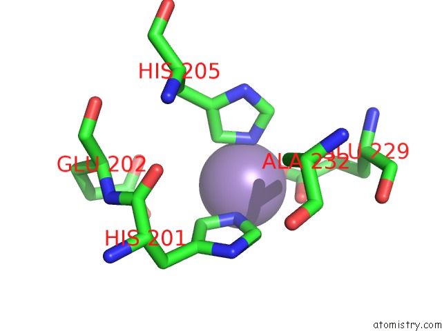
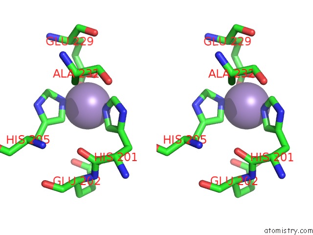
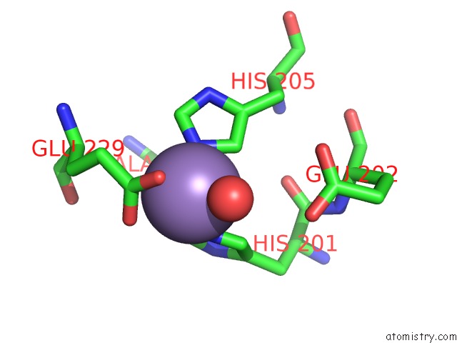
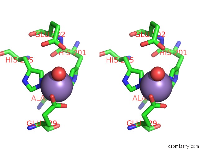
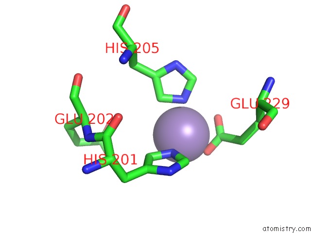
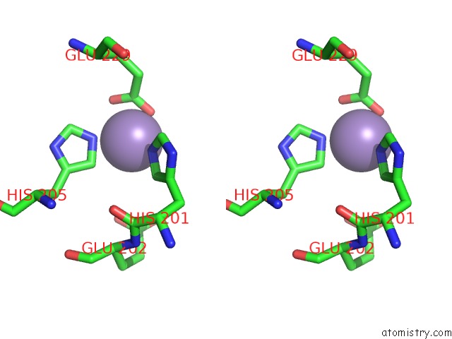
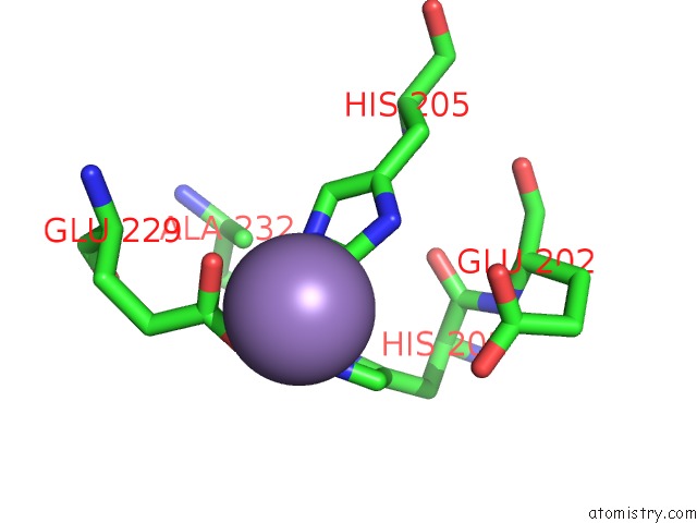
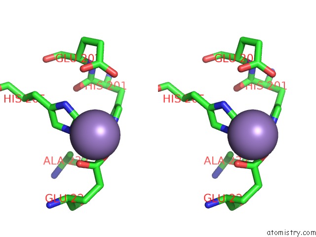
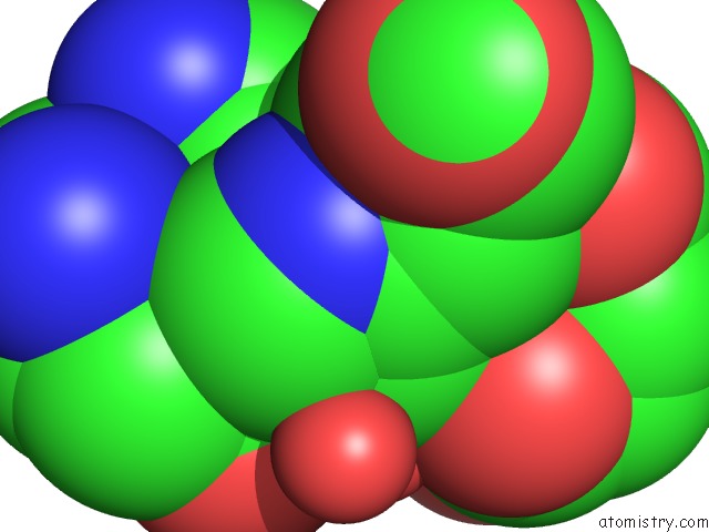
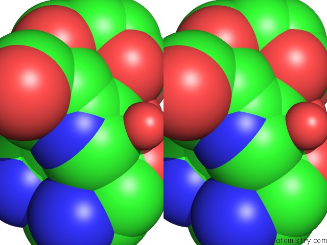
Manganese binding site 1 out of 8 in 6sna
Go back to
Manganese binding site 1 out
of 8 in the Crystal Structure of Antirestriction Ardc Protein From R388 Plasmid. Mn(II)-Bound Structure.
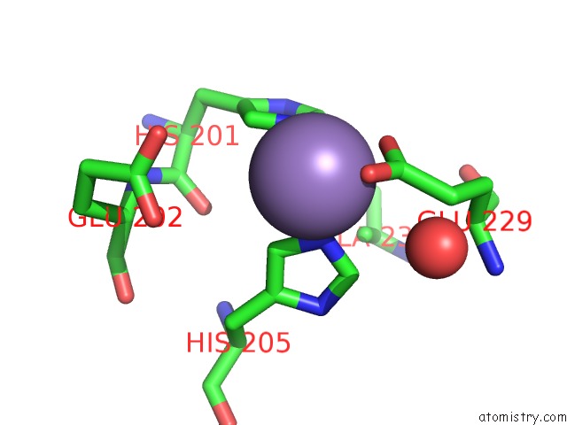
Mono view
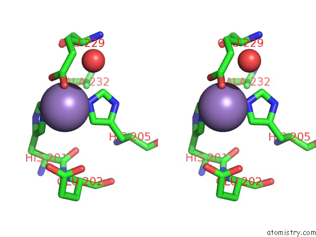
Stereo pair view
Mono view
Stereo pair view
A full contact list of Manganese with other atoms in the Mn binding
site number 1 of Crystal Structure of Antirestriction Ardc Protein From R388 Plasmid. Mn(II)-Bound Structure. within 5.0Å range:
|
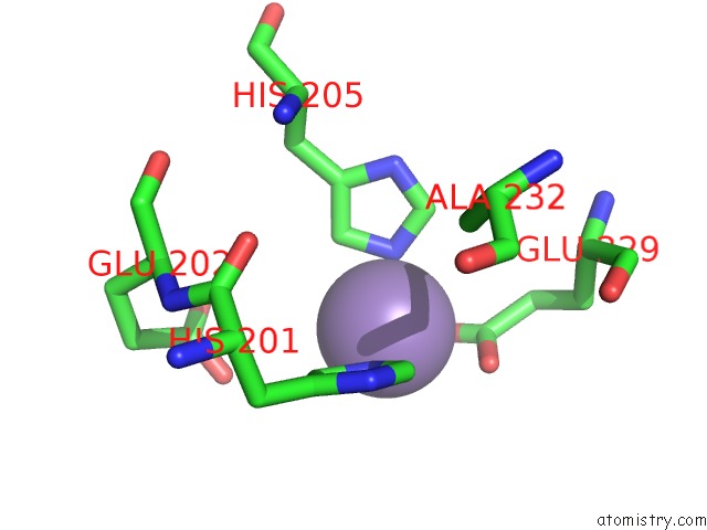
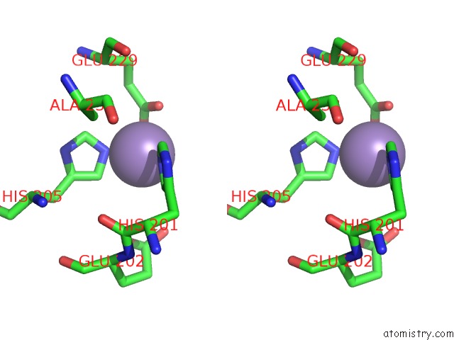
Manganese binding site 2 out of 8 in 6sna
Go back to
Manganese binding site 2 out
of 8 in the Crystal Structure of Antirestriction Ardc Protein From R388 Plasmid. Mn(II)-Bound Structure.
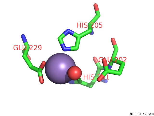
Mono view
Stereo pair view
Mono view
Stereo pair view
A full contact list of Manganese with other atoms in the Mn binding
site number 2 of Crystal Structure of Antirestriction Ardc Protein From R388 Plasmid. Mn(II)-Bound Structure. within 5.0Å range:
|
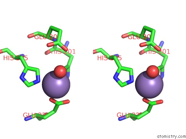
Manganese binding site 3 out of 8 in 6sna
Go back to
Manganese binding site 3 out
of 8 in the Crystal Structure of Antirestriction Ardc Protein From R388 Plasmid. Mn(II)-Bound Structure.
Mono view
Stereo pair view
Mono view
Stereo pair view
A full contact list of Manganese with other atoms in the Mn binding
site number 3 of Crystal Structure of Antirestriction Ardc Protein From R388 Plasmid. Mn(II)-Bound Structure. within 5.0Å range:
|
Manganese binding site 4 out of 8 in 6sna
Go back to
Manganese binding site 4 out
of 8 in the Crystal Structure of Antirestriction Ardc Protein From R388 Plasmid. Mn(II)-Bound Structure.
Mono view
Stereo pair view
Mono view
Stereo pair view
A full contact list of Manganese with other atoms in the Mn binding
site number 4 of Crystal Structure of Antirestriction Ardc Protein From R388 Plasmid. Mn(II)-Bound Structure. within 5.0Å range:
|
Manganese binding site 5 out of 8 in 6sna
Go back to
Manganese binding site 5 out
of 8 in the Crystal Structure of Antirestriction Ardc Protein From R388 Plasmid. Mn(II)-Bound Structure.
Mono view
Stereo pair view
Mono view
Stereo pair view
A full contact list of Manganese with other atoms in the Mn binding
site number 5 of Crystal Structure of Antirestriction Ardc Protein From R388 Plasmid. Mn(II)-Bound Structure. within 5.0Å range:
|
Manganese binding site 6 out of 8 in 6sna
Go back to
Manganese binding site 6 out
of 8 in the Crystal Structure of Antirestriction Ardc Protein From R388 Plasmid. Mn(II)-Bound Structure.
Mono view
Stereo pair view
Mono view
Stereo pair view
A full contact list of Manganese with other atoms in the Mn binding
site number 6 of Crystal Structure of Antirestriction Ardc Protein From R388 Plasmid. Mn(II)-Bound Structure. within 5.0Å range:
|
Manganese binding site 7 out of 8 in 6sna
Go back to
Manganese binding site 7 out
of 8 in the Crystal Structure of Antirestriction Ardc Protein From R388 Plasmid. Mn(II)-Bound Structure.
Mono view
Stereo pair view
Mono view
Stereo pair view
A full contact list of Manganese with other atoms in the Mn binding
site number 7 of Crystal Structure of Antirestriction Ardc Protein From R388 Plasmid. Mn(II)-Bound Structure. within 5.0Å range:
|
Manganese binding site 8 out of 8 in 6sna
Go back to
Manganese binding site 8 out
of 8 in the Crystal Structure of Antirestriction Ardc Protein From R388 Plasmid. Mn(II)-Bound Structure.
Mono view
Stereo pair view
Mono view
Stereo pair view
A full contact list of Manganese with other atoms in the Mn binding
site number 8 of Crystal Structure of Antirestriction Ardc Protein From R388 Plasmid. Mn(II)-Bound Structure. within 5.0Å range:
|
Reference:
L.Gonzalez-Montes,
G.Moncalian.
Crystal Structure of Antirestriction Ardc Protein From R388 Plasmid. To Be Published.
Page generated: Sun Oct 6 07:08:26 2024
Last articles
I in 8IKPI in 8IKS
I in 8ING
I in 8IKB
I in 8H3Z
I in 8H5L
I in 8II3
I in 8II1
I in 8IG1
I in 8GYR