Manganese »
PDB 6q9p-6qv8 »
6qeh »
Manganese in PDB 6qeh: Crystal Structure of Human Methionine Aminopeptidase-2 in Complex with An Inhibitor 5-Chloro-Quinolin-8-Ol
Enzymatic activity of Crystal Structure of Human Methionine Aminopeptidase-2 in Complex with An Inhibitor 5-Chloro-Quinolin-8-Ol
All present enzymatic activity of Crystal Structure of Human Methionine Aminopeptidase-2 in Complex with An Inhibitor 5-Chloro-Quinolin-8-Ol:
3.4.11.18;
3.4.11.18;
Protein crystallography data
The structure of Crystal Structure of Human Methionine Aminopeptidase-2 in Complex with An Inhibitor 5-Chloro-Quinolin-8-Ol, PDB code: 6qeh
was solved by
D.Musil,
T.Heinrich,
M.Lehmann,
with X-Ray Crystallography technique. A brief refinement statistics is given in the table below:
| Resolution Low / High (Å) | 19.01 / 2.17 |
| Space group | C 2 2 21 |
| Cell size a, b, c (Å), α, β, γ (°) | 90.060, 100.260, 100.340, 90.00, 90.00, 90.00 |
| R / Rfree (%) | 18.4 / 22 |
Other elements in 6qeh:
The structure of Crystal Structure of Human Methionine Aminopeptidase-2 in Complex with An Inhibitor 5-Chloro-Quinolin-8-Ol also contains other interesting chemical elements:
| Chlorine | (Cl) | 1 atom |
Manganese Binding Sites:
The binding sites of Manganese atom in the Crystal Structure of Human Methionine Aminopeptidase-2 in Complex with An Inhibitor 5-Chloro-Quinolin-8-Ol
(pdb code 6qeh). This binding sites where shown within
5.0 Angstroms radius around Manganese atom.
In total 2 binding sites of Manganese where determined in the Crystal Structure of Human Methionine Aminopeptidase-2 in Complex with An Inhibitor 5-Chloro-Quinolin-8-Ol, PDB code: 6qeh:
Jump to Manganese binding site number: 1; 2;
In total 2 binding sites of Manganese where determined in the Crystal Structure of Human Methionine Aminopeptidase-2 in Complex with An Inhibitor 5-Chloro-Quinolin-8-Ol, PDB code: 6qeh:
Jump to Manganese binding site number: 1; 2;
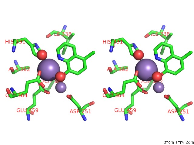
Manganese binding site 1 out of 2 in 6qeh
Go back to
Manganese binding site 1 out
of 2 in the Crystal Structure of Human Methionine Aminopeptidase-2 in Complex with An Inhibitor 5-Chloro-Quinolin-8-Ol

Mono view
Stereo pair view
Mono view
Stereo pair view
A full contact list of Manganese with other atoms in the Mn binding
site number 1 of Crystal Structure of Human Methionine Aminopeptidase-2 in Complex with An Inhibitor 5-Chloro-Quinolin-8-Ol within 5.0Å range:
|
Manganese binding site 2 out of 2 in 6qeh
Go back to
Manganese binding site 2 out
of 2 in the Crystal Structure of Human Methionine Aminopeptidase-2 in Complex with An Inhibitor 5-Chloro-Quinolin-8-Ol

Mono view

Stereo pair view
Mono view
Stereo pair view
A full contact list of Manganese with other atoms in the Mn binding
site number 2 of Crystal Structure of Human Methionine Aminopeptidase-2 in Complex with An Inhibitor 5-Chloro-Quinolin-8-Ol within 5.0Å range:
|
Reference:
T.Heinrich,
J.Seenisamy,
B.Blume,
J.Bomke,
M.Calderini,
U.Eckert,
M.Friese-Hamim,
R.Kohl,
M.Lehmann,
B.Leuthner,
D.Musil,
F.Rohdich,
F.T.Zenke.
Discovery and Structure-Based Optimization of Next-Generation Reversible Methionine Aminopeptidase-2 (Metap-2) Inhibitors. J.Med.Chem. V. 62 5025 2019.
ISSN: ISSN 0022-2623
PubMed: 30939017
DOI: 10.1021/ACS.JMEDCHEM.9B00041
Page generated: Sun Oct 6 05:56:23 2024
ISSN: ISSN 0022-2623
PubMed: 30939017
DOI: 10.1021/ACS.JMEDCHEM.9B00041
Last articles
Mg in 4W5OMg in 4W5J
Mg in 4W5N
Mg in 4V2I
Mg in 4V3R
Mg in 4V26
Mg in 4V2G
Mg in 4V1T
Mg in 4V25
Mg in 4V1V