Manganese »
PDB 6f4q-6fxr »
6fm4 »
Manganese in PDB 6fm4: The Crystal Structure of S. Aureus Gyrase Complex with Id-130 and Dna
Enzymatic activity of The Crystal Structure of S. Aureus Gyrase Complex with Id-130 and Dna
All present enzymatic activity of The Crystal Structure of S. Aureus Gyrase Complex with Id-130 and Dna:
5.99.1.3;
5.99.1.3;
Protein crystallography data
The structure of The Crystal Structure of S. Aureus Gyrase Complex with Id-130 and Dna, PDB code: 6fm4
was solved by
R.Ombrato,
B.Garofalo,
G.Mangano,
F.Mancini,
with X-Ray Crystallography technique. A brief refinement statistics is given in the table below:
| Resolution Low / High (Å) | 43.05 / 2.70 |
| Space group | P 61 |
| Cell size a, b, c (Å), α, β, γ (°) | 92.951, 92.951, 407.030, 90.00, 90.00, 120.00 |
| R / Rfree (%) | 22.5 / 30 |
Manganese Binding Sites:
The binding sites of Manganese atom in the The Crystal Structure of S. Aureus Gyrase Complex with Id-130 and Dna
(pdb code 6fm4). This binding sites where shown within
5.0 Angstroms radius around Manganese atom.
In total 3 binding sites of Manganese where determined in the The Crystal Structure of S. Aureus Gyrase Complex with Id-130 and Dna, PDB code: 6fm4:
Jump to Manganese binding site number: 1; 2; 3;
In total 3 binding sites of Manganese where determined in the The Crystal Structure of S. Aureus Gyrase Complex with Id-130 and Dna, PDB code: 6fm4:
Jump to Manganese binding site number: 1; 2; 3;
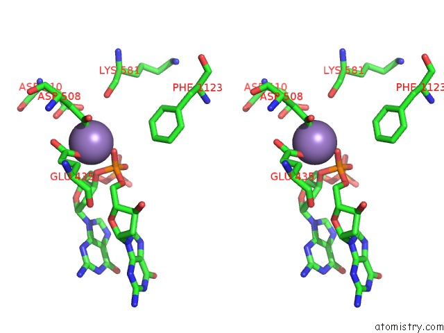
Manganese binding site 1 out of 3 in 6fm4
Go back to
Manganese binding site 1 out
of 3 in the The Crystal Structure of S. Aureus Gyrase Complex with Id-130 and Dna

Mono view
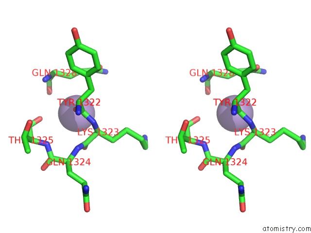
Stereo pair view
Mono view
Stereo pair view
A full contact list of Manganese with other atoms in the Mn binding
site number 1 of The Crystal Structure of S. Aureus Gyrase Complex with Id-130 and Dna within 5.0Å range:
|
Manganese binding site 2 out of 3 in 6fm4
Go back to
Manganese binding site 2 out
of 3 in the The Crystal Structure of S. Aureus Gyrase Complex with Id-130 and Dna
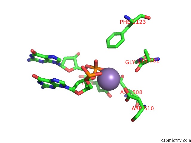
Mono view
Stereo pair view
Mono view
Stereo pair view
A full contact list of Manganese with other atoms in the Mn binding
site number 2 of The Crystal Structure of S. Aureus Gyrase Complex with Id-130 and Dna within 5.0Å range:
|
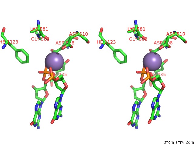
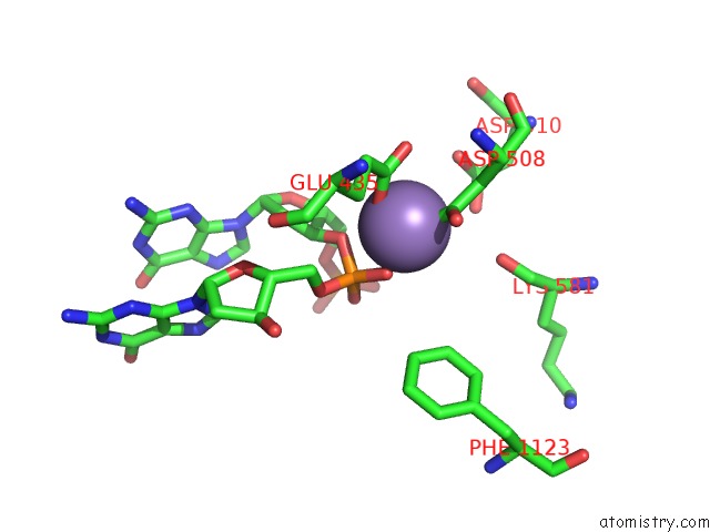
Manganese binding site 3 out of 3 in 6fm4
Go back to
Manganese binding site 3 out
of 3 in the The Crystal Structure of S. Aureus Gyrase Complex with Id-130 and Dna
Mono view
Stereo pair view
Mono view
Stereo pair view
A full contact list of Manganese with other atoms in the Mn binding
site number 3 of The Crystal Structure of S. Aureus Gyrase Complex with Id-130 and Dna within 5.0Å range:
|
Reference:
G.Magaro,
F.Prati,
B.Garofalo,
G.Corso,
G.Furlotti,
C.Apicella,
G.Mangano,
N.D'atanasio,
D.Robinson,
F.P.Di Giorgio,
R.Ombrato.
Virtual Screening Approach and Investigation of Structure-Activity Relationships to Discover Novel Bacterial Topoisomerase Inhibitors Targeting Gram-Positive and Gram-Negative Pathogens. J.Med.Chem. V. 62 7445 2019.
ISSN: ISSN 0022-2623
PubMed: 31276392
DOI: 10.1021/ACS.JMEDCHEM.9B00394
Page generated: Sun Oct 6 04:37:15 2024
ISSN: ISSN 0022-2623
PubMed: 31276392
DOI: 10.1021/ACS.JMEDCHEM.9B00394
Last articles
K in 6D7HK in 6DE8
K in 6D1P
K in 6D2J
K in 6CW8
K in 6CSS
K in 6CST
K in 6CZ3
K in 6CSR
K in 6CSQ