Manganese »
PDB 5z2k-6a9u »
5zqv »
Manganese in PDB 5zqv: Crystal Structure of Protein Phosphate 1 Complexed with PP1 Binding Domain of Gm
Enzymatic activity of Crystal Structure of Protein Phosphate 1 Complexed with PP1 Binding Domain of Gm
All present enzymatic activity of Crystal Structure of Protein Phosphate 1 Complexed with PP1 Binding Domain of Gm:
3.1.3.16;
3.1.3.16;
Protein crystallography data
The structure of Crystal Structure of Protein Phosphate 1 Complexed with PP1 Binding Domain of Gm, PDB code: 5zqv
was solved by
J.Yu,
S.Xiang,
with X-Ray Crystallography technique. A brief refinement statistics is given in the table below:
| Resolution Low / High (Å) | 33.29 / 2.95 |
| Space group | P 41 |
| Cell size a, b, c (Å), α, β, γ (°) | 111.973, 111.973, 195.299, 90.00, 90.00, 90.00 |
| R / Rfree (%) | 25.2 / 29.7 |
Manganese Binding Sites:
The binding sites of Manganese atom in the Crystal Structure of Protein Phosphate 1 Complexed with PP1 Binding Domain of Gm
(pdb code 5zqv). This binding sites where shown within
5.0 Angstroms radius around Manganese atom.
In total 4 binding sites of Manganese where determined in the Crystal Structure of Protein Phosphate 1 Complexed with PP1 Binding Domain of Gm, PDB code: 5zqv:
Jump to Manganese binding site number: 1; 2; 3; 4;
In total 4 binding sites of Manganese where determined in the Crystal Structure of Protein Phosphate 1 Complexed with PP1 Binding Domain of Gm, PDB code: 5zqv:
Jump to Manganese binding site number: 1; 2; 3; 4;
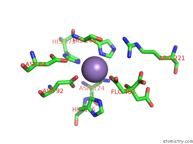
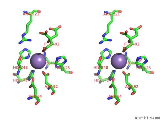
Manganese binding site 1 out of 4 in 5zqv
Go back to
Manganese binding site 1 out
of 4 in the Crystal Structure of Protein Phosphate 1 Complexed with PP1 Binding Domain of Gm
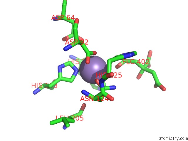
Mono view
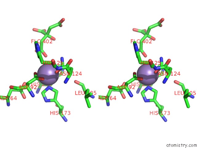
Stereo pair view
Mono view
Stereo pair view
A full contact list of Manganese with other atoms in the Mn binding
site number 1 of Crystal Structure of Protein Phosphate 1 Complexed with PP1 Binding Domain of Gm within 5.0Å range:
|
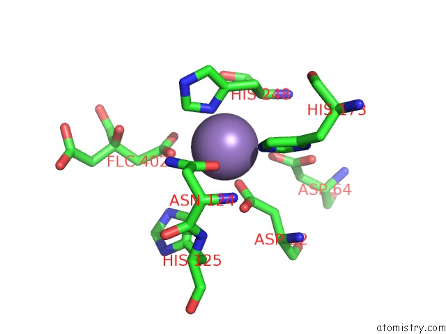
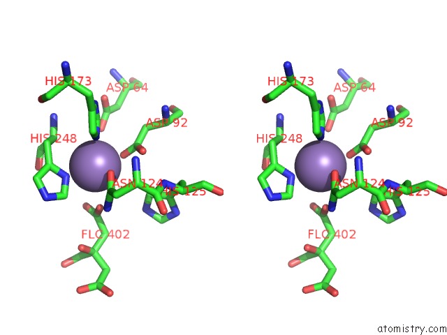
Manganese binding site 2 out of 4 in 5zqv
Go back to
Manganese binding site 2 out
of 4 in the Crystal Structure of Protein Phosphate 1 Complexed with PP1 Binding Domain of Gm
Mono view
Stereo pair view
Mono view
Stereo pair view
A full contact list of Manganese with other atoms in the Mn binding
site number 2 of Crystal Structure of Protein Phosphate 1 Complexed with PP1 Binding Domain of Gm within 5.0Å range:
|
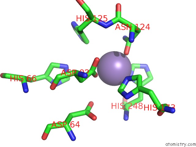
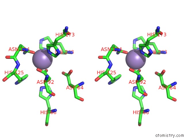
Manganese binding site 3 out of 4 in 5zqv
Go back to
Manganese binding site 3 out
of 4 in the Crystal Structure of Protein Phosphate 1 Complexed with PP1 Binding Domain of Gm
Mono view
Stereo pair view
Mono view
Stereo pair view
A full contact list of Manganese with other atoms in the Mn binding
site number 3 of Crystal Structure of Protein Phosphate 1 Complexed with PP1 Binding Domain of Gm within 5.0Å range:
|
Manganese binding site 4 out of 4 in 5zqv
Go back to
Manganese binding site 4 out
of 4 in the Crystal Structure of Protein Phosphate 1 Complexed with PP1 Binding Domain of Gm
Mono view
Stereo pair view
Mono view
Stereo pair view
A full contact list of Manganese with other atoms in the Mn binding
site number 4 of Crystal Structure of Protein Phosphate 1 Complexed with PP1 Binding Domain of Gm within 5.0Å range:
|
Reference:
J.Yu,
T.Deng,
S.Xiang.
Structural Basis For Protein Phosphatase 1 Recruitment By Glycogen-Targeting Subunits. Febs J. V. 285 4646 2018.
ISSN: ISSN 1742-4658
PubMed: 30422398
DOI: 10.1111/FEBS.14699
Page generated: Sun Oct 6 03:44:33 2024
ISSN: ISSN 1742-4658
PubMed: 30422398
DOI: 10.1111/FEBS.14699
Last articles
I in 5ZKZI in 5ZK7
I in 5ZIW
I in 5ZGS
I in 5ZII
I in 5ZH9
I in 5ZH0
I in 5YGU
I in 5ZF3
I in 5WLH