Manganese »
PDB 5z2k-6a9u »
5zqn »
Manganese in PDB 5zqn: Crystal Structure of Mycobacterium Tuberculosis Hisb in Complex with A Ligand
Enzymatic activity of Crystal Structure of Mycobacterium Tuberculosis Hisb in Complex with A Ligand
All present enzymatic activity of Crystal Structure of Mycobacterium Tuberculosis Hisb in Complex with A Ligand:
4.2.1.19;
4.2.1.19;
Protein crystallography data
The structure of Crystal Structure of Mycobacterium Tuberculosis Hisb in Complex with A Ligand, PDB code: 5zqn
was solved by
D.Kumar,
R.K.Pal,
B.K.Biswal,
with X-Ray Crystallography technique. A brief refinement statistics is given in the table below:
| Resolution Low / High (Å) | 28.19 / 1.80 |
| Space group | P 4 3 2 |
| Cell size a, b, c (Å), α, β, γ (°) | 112.715, 112.715, 112.715, 90.00, 90.00, 90.00 |
| R / Rfree (%) | 15.4 / 18.7 |
Other elements in 5zqn:
The structure of Crystal Structure of Mycobacterium Tuberculosis Hisb in Complex with A Ligand also contains other interesting chemical elements:
| Chlorine | (Cl) | 1 atom |
Manganese Binding Sites:
The binding sites of Manganese atom in the Crystal Structure of Mycobacterium Tuberculosis Hisb in Complex with A Ligand
(pdb code 5zqn). This binding sites where shown within
5.0 Angstroms radius around Manganese atom.
In total 2 binding sites of Manganese where determined in the Crystal Structure of Mycobacterium Tuberculosis Hisb in Complex with A Ligand, PDB code: 5zqn:
Jump to Manganese binding site number: 1; 2;
In total 2 binding sites of Manganese where determined in the Crystal Structure of Mycobacterium Tuberculosis Hisb in Complex with A Ligand, PDB code: 5zqn:
Jump to Manganese binding site number: 1; 2;
Manganese binding site 1 out of 2 in 5zqn
Go back to
Manganese binding site 1 out
of 2 in the Crystal Structure of Mycobacterium Tuberculosis Hisb in Complex with A Ligand
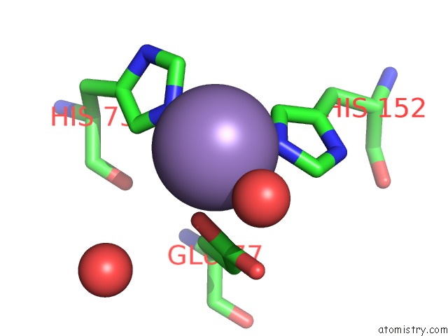
Mono view
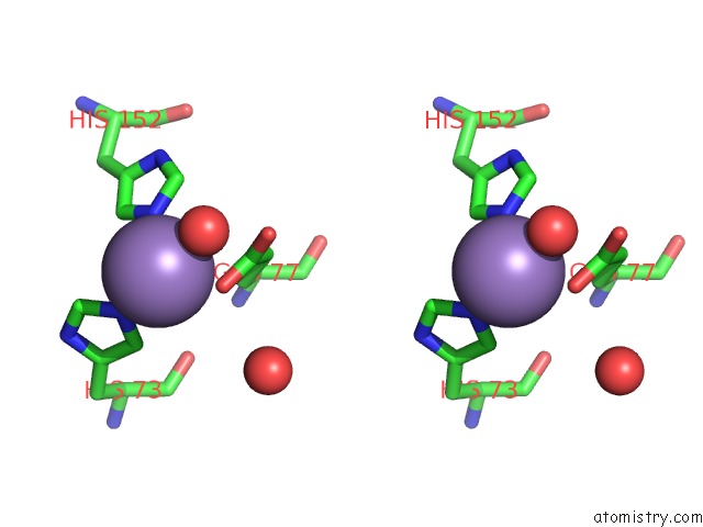
Stereo pair view
Mono view
Stereo pair view
A full contact list of Manganese with other atoms in the Mn binding
site number 1 of Crystal Structure of Mycobacterium Tuberculosis Hisb in Complex with A Ligand within 5.0Å range:
|
Manganese binding site 2 out of 2 in 5zqn
Go back to
Manganese binding site 2 out
of 2 in the Crystal Structure of Mycobacterium Tuberculosis Hisb in Complex with A Ligand
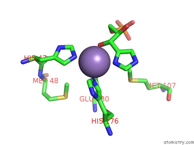
Mono view
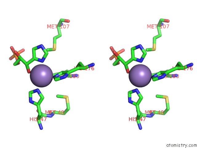
Stereo pair view
Mono view
Stereo pair view
A full contact list of Manganese with other atoms in the Mn binding
site number 2 of Crystal Structure of Mycobacterium Tuberculosis Hisb in Complex with A Ligand within 5.0Å range:
|
Reference:
D.Kumar,
R.K.Pal,
B.K.Biswal.
Crystal Structure of Mycobacterium Tuberculosis Hisb in Complex with A Ligand To Be Published.
Page generated: Sun Oct 6 03:44:01 2024
Last articles
K in 3MIJK in 3MD7
K in 3M62
K in 3M63
K in 3LNM
K in 3LUT
K in 3LIB
K in 3LQX
K in 3LLP
K in 3LMS