Manganese »
PDB 5z2k-6a9u »
5zeh »
Manganese in PDB 5zeh: Crystal Structure of Entamoeba Histolytica Arginase in Complex with L- Ornithine at 2.35 A
Enzymatic activity of Crystal Structure of Entamoeba Histolytica Arginase in Complex with L- Ornithine at 2.35 A
All present enzymatic activity of Crystal Structure of Entamoeba Histolytica Arginase in Complex with L- Ornithine at 2.35 A:
3.5.3.1;
3.5.3.1;
Protein crystallography data
The structure of Crystal Structure of Entamoeba Histolytica Arginase in Complex with L- Ornithine at 2.35 A, PDB code: 5zeh
was solved by
A.Malik,
V.Dalal,
S.Ankri,
S.Tomar,
with X-Ray Crystallography technique. A brief refinement statistics is given in the table below:
| Resolution Low / High (Å) | 78.68 / 2.36 |
| Space group | I 21 21 21 |
| Cell size a, b, c (Å), α, β, γ (°) | 87.605, 97.494, 133.255, 90.00, 90.00, 90.00 |
| R / Rfree (%) | 21.9 / 26.2 |
Manganese Binding Sites:
The binding sites of Manganese atom in the Crystal Structure of Entamoeba Histolytica Arginase in Complex with L- Ornithine at 2.35 A
(pdb code 5zeh). This binding sites where shown within
5.0 Angstroms radius around Manganese atom.
In total 4 binding sites of Manganese where determined in the Crystal Structure of Entamoeba Histolytica Arginase in Complex with L- Ornithine at 2.35 A, PDB code: 5zeh:
Jump to Manganese binding site number: 1; 2; 3; 4;
In total 4 binding sites of Manganese where determined in the Crystal Structure of Entamoeba Histolytica Arginase in Complex with L- Ornithine at 2.35 A, PDB code: 5zeh:
Jump to Manganese binding site number: 1; 2; 3; 4;
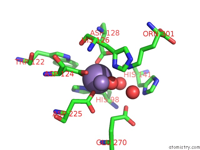
Manganese binding site 1 out of 4 in 5zeh
Go back to
Manganese binding site 1 out
of 4 in the Crystal Structure of Entamoeba Histolytica Arginase in Complex with L- Ornithine at 2.35 A
Mono view

Stereo pair view
Mono view
Stereo pair view
A full contact list of Manganese with other atoms in the Mn binding
site number 1 of Crystal Structure of Entamoeba Histolytica Arginase in Complex with L- Ornithine at 2.35 A within 5.0Å range:
|
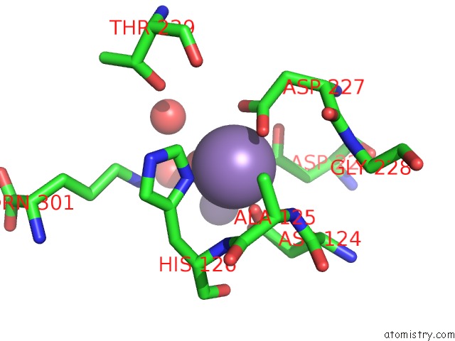
Manganese binding site 2 out of 4 in 5zeh
Go back to
Manganese binding site 2 out
of 4 in the Crystal Structure of Entamoeba Histolytica Arginase in Complex with L- Ornithine at 2.35 A
Mono view

Stereo pair view
Mono view
Stereo pair view
A full contact list of Manganese with other atoms in the Mn binding
site number 2 of Crystal Structure of Entamoeba Histolytica Arginase in Complex with L- Ornithine at 2.35 A within 5.0Å range:
|
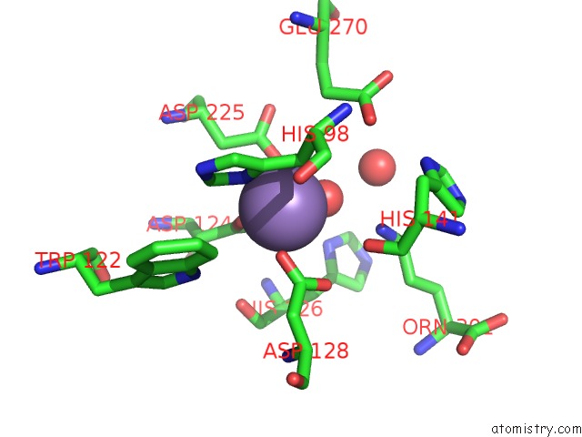
Manganese binding site 3 out of 4 in 5zeh
Go back to
Manganese binding site 3 out
of 4 in the Crystal Structure of Entamoeba Histolytica Arginase in Complex with L- Ornithine at 2.35 A
Mono view

Stereo pair view
Mono view
Stereo pair view
A full contact list of Manganese with other atoms in the Mn binding
site number 3 of Crystal Structure of Entamoeba Histolytica Arginase in Complex with L- Ornithine at 2.35 A within 5.0Å range:
|
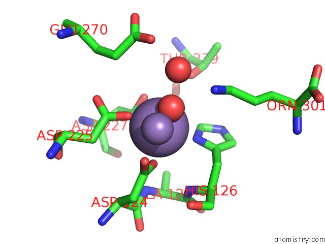
Manganese binding site 4 out of 4 in 5zeh
Go back to
Manganese binding site 4 out
of 4 in the Crystal Structure of Entamoeba Histolytica Arginase in Complex with L- Ornithine at 2.35 A
Mono view

Stereo pair view
Mono view
Stereo pair view
A full contact list of Manganese with other atoms in the Mn binding
site number 4 of Crystal Structure of Entamoeba Histolytica Arginase in Complex with L- Ornithine at 2.35 A within 5.0Å range:
|
Reference:
A.Malik,
V.Dalal,
S.Ankri,
S.Tomar.
Structural Insights Into Entamoeba Histolytica Arginase and Structure-Based Identification of Novel Non-Amino Acid Based Inhibitors As Potential Antiamoebic Molecules. Febs J. V. 286 4135 2019.
ISSN: ISSN 1742-464X
PubMed: 31199070
DOI: 10.1111/FEBS.14960
Page generated: Sun Oct 6 03:41:02 2024
ISSN: ISSN 1742-464X
PubMed: 31199070
DOI: 10.1111/FEBS.14960
Last articles
I in 6AXXI in 6AXW
I in 6AXT
I in 6AXV
I in 6AXS
I in 6AN0
I in 6AXR
I in 5ZBA
I in 6A4Y
I in 5ZRF