Manganese »
PDB 5vqn-5wg9 »
5vr0 »
Manganese in PDB 5vr0: Crystal Structure of Glucose Isomerase From Streptomyces Rubiginosus
Enzymatic activity of Crystal Structure of Glucose Isomerase From Streptomyces Rubiginosus
All present enzymatic activity of Crystal Structure of Glucose Isomerase From Streptomyces Rubiginosus:
5.3.1.5;
5.3.1.5;
Protein crystallography data
The structure of Crystal Structure of Glucose Isomerase From Streptomyces Rubiginosus, PDB code: 5vr0
was solved by
D.Borek,
Z.Otwinowski,
with X-Ray Crystallography technique. A brief refinement statistics is given in the table below:
| Resolution Low / High (Å) | 38.90 / 1.70 |
| Space group | I 2 2 2 |
| Cell size a, b, c (Å), α, β, γ (°) | 92.854, 98.061, 102.279, 90.00, 90.00, 90.00 |
| R / Rfree (%) | 11.9 / 14.2 |
Other elements in 5vr0:
The structure of Crystal Structure of Glucose Isomerase From Streptomyces Rubiginosus also contains other interesting chemical elements:
| Chlorine | (Cl) | 12 atoms |
| Calcium | (Ca) | 3 atoms |
Manganese Binding Sites:
The binding sites of Manganese atom in the Crystal Structure of Glucose Isomerase From Streptomyces Rubiginosus
(pdb code 5vr0). This binding sites where shown within
5.0 Angstroms radius around Manganese atom.
In total 2 binding sites of Manganese where determined in the Crystal Structure of Glucose Isomerase From Streptomyces Rubiginosus, PDB code: 5vr0:
Jump to Manganese binding site number: 1; 2;
In total 2 binding sites of Manganese where determined in the Crystal Structure of Glucose Isomerase From Streptomyces Rubiginosus, PDB code: 5vr0:
Jump to Manganese binding site number: 1; 2;
Manganese binding site 1 out of 2 in 5vr0
Go back to
Manganese binding site 1 out
of 2 in the Crystal Structure of Glucose Isomerase From Streptomyces Rubiginosus
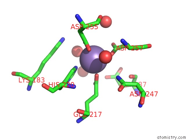
Mono view

Stereo pair view
Mono view
Stereo pair view
A full contact list of Manganese with other atoms in the Mn binding
site number 1 of Crystal Structure of Glucose Isomerase From Streptomyces Rubiginosus within 5.0Å range:
|
Manganese binding site 2 out of 2 in 5vr0
Go back to
Manganese binding site 2 out
of 2 in the Crystal Structure of Glucose Isomerase From Streptomyces Rubiginosus
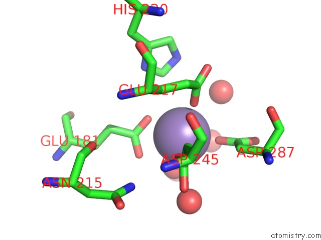
Mono view
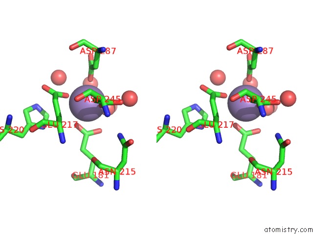
Stereo pair view
Mono view
Stereo pair view
A full contact list of Manganese with other atoms in the Mn binding
site number 2 of Crystal Structure of Glucose Isomerase From Streptomyces Rubiginosus within 5.0Å range:
|
Reference:
D.Borek,
R.Bromberg,
J.Hattne,
Z.Otwinowski.
Real-Space Analysis of Radiation-Induced Specific Changes with Independent Component Analysis. J Synchrotron Radiat V. 25 451 2018.
ISSN: ESSN 1600-5775
PubMed: 29488925
DOI: 10.1107/S1600577517018148
Page generated: Sun Oct 6 03:13:48 2024
ISSN: ESSN 1600-5775
PubMed: 29488925
DOI: 10.1107/S1600577517018148
Last articles
Zn in 9MJ5Zn in 9HNW
Zn in 9G0L
Zn in 9FNE
Zn in 9DZN
Zn in 9E0I
Zn in 9D32
Zn in 9DAK
Zn in 8ZXC
Zn in 8ZUF