Manganese »
PDB 5svc-5uqn »
5u9z »
Manganese in PDB 5u9z: Phosphoglycerol Transferase Gach From Streptococcus Pyogenes
Protein crystallography data
The structure of Phosphoglycerol Transferase Gach From Streptococcus Pyogenes, PDB code: 5u9z
was solved by
R.J.Edgar,
N.Korotkova,
K.V.Korotkov,
with X-Ray Crystallography technique. A brief refinement statistics is given in the table below:
| Resolution Low / High (Å) | 49.51 / 2.00 |
| Space group | P 21 21 21 |
| Cell size a, b, c (Å), α, β, γ (°) | 68.870, 78.170, 172.660, 90.00, 90.00, 90.00 |
| R / Rfree (%) | 21.5 / 24 |
Manganese Binding Sites:
The binding sites of Manganese atom in the Phosphoglycerol Transferase Gach From Streptococcus Pyogenes
(pdb code 5u9z). This binding sites where shown within
5.0 Angstroms radius around Manganese atom.
In total 2 binding sites of Manganese where determined in the Phosphoglycerol Transferase Gach From Streptococcus Pyogenes, PDB code: 5u9z:
Jump to Manganese binding site number: 1; 2;
In total 2 binding sites of Manganese where determined in the Phosphoglycerol Transferase Gach From Streptococcus Pyogenes, PDB code: 5u9z:
Jump to Manganese binding site number: 1; 2;
Manganese binding site 1 out of 2 in 5u9z
Go back to
Manganese binding site 1 out
of 2 in the Phosphoglycerol Transferase Gach From Streptococcus Pyogenes
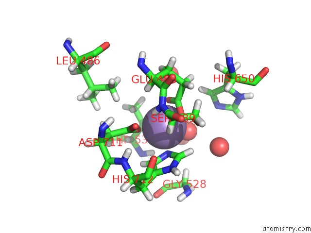
Mono view
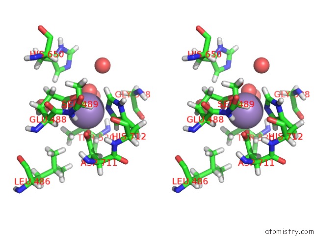
Stereo pair view
Mono view
Stereo pair view
A full contact list of Manganese with other atoms in the Mn binding
site number 1 of Phosphoglycerol Transferase Gach From Streptococcus Pyogenes within 5.0Å range:
|
Manganese binding site 2 out of 2 in 5u9z
Go back to
Manganese binding site 2 out
of 2 in the Phosphoglycerol Transferase Gach From Streptococcus Pyogenes
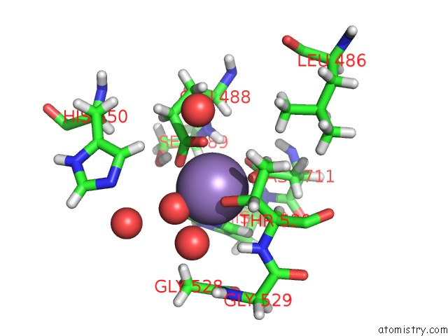
Mono view
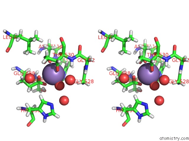
Stereo pair view
Mono view
Stereo pair view
A full contact list of Manganese with other atoms in the Mn binding
site number 2 of Phosphoglycerol Transferase Gach From Streptococcus Pyogenes within 5.0Å range:
|
Reference:
R.J.Edgar,
V.P.Van Hensbergen,
A.Ruda,
A.G.Turner,
P.Deng,
Y.Le Breton,
N.M.El-Sayed,
A.T.Belew,
K.S.Mciver,
A.G.Mcewan,
A.J.Morris,
G.Lambeau,
M.J.Walker,
J.S.Rush,
K.V.Korotkov,
G.Widmalm,
N.M.Van Sorge,
N.Korotkova.
Discovery of Glycerol Phosphate Modification on Streptococcal Rhamnose Polysaccharides. Nat.Chem.Biol. V. 15 463 2019.
ISSN: ESSN 1552-4469
PubMed: 30936502
DOI: 10.1038/S41589-019-0251-4
Page generated: Sun Oct 6 03:02:56 2024
ISSN: ESSN 1552-4469
PubMed: 30936502
DOI: 10.1038/S41589-019-0251-4
Last articles
I in 3WN5I in 3WYX
I in 3WGW
I in 3WD6
I in 3WB5
I in 3W31
I in 3WB4
I in 3W1N
I in 3W0F
I in 3W2Z