Manganese »
PDB 5lyx-5nh9 »
5n2g »
Manganese in PDB 5n2g: Structure of the E9 Dna Polymerase From Vaccinia Virus in Complex with Manganese
Enzymatic activity of Structure of the E9 Dna Polymerase From Vaccinia Virus in Complex with Manganese
All present enzymatic activity of Structure of the E9 Dna Polymerase From Vaccinia Virus in Complex with Manganese:
2.7.7.7;
2.7.7.7;
Protein crystallography data
The structure of Structure of the E9 Dna Polymerase From Vaccinia Virus in Complex with Manganese, PDB code: 5n2g
was solved by
N.Tarbouriech,
W.P.Burmeister,
F.Iseni,
with X-Ray Crystallography technique. A brief refinement statistics is given in the table below:
| Resolution Low / High (Å) | 46.00 / 2.78 |
| Space group | P 31 2 1 |
| Cell size a, b, c (Å), α, β, γ (°) | 134.007, 134.007, 230.189, 90.00, 90.00, 120.00 |
| R / Rfree (%) | 18.3 / 22.7 |
Manganese Binding Sites:
The binding sites of Manganese atom in the Structure of the E9 Dna Polymerase From Vaccinia Virus in Complex with Manganese
(pdb code 5n2g). This binding sites where shown within
5.0 Angstroms radius around Manganese atom.
In total 4 binding sites of Manganese where determined in the Structure of the E9 Dna Polymerase From Vaccinia Virus in Complex with Manganese, PDB code: 5n2g:
Jump to Manganese binding site number: 1; 2; 3; 4;
In total 4 binding sites of Manganese where determined in the Structure of the E9 Dna Polymerase From Vaccinia Virus in Complex with Manganese, PDB code: 5n2g:
Jump to Manganese binding site number: 1; 2; 3; 4;
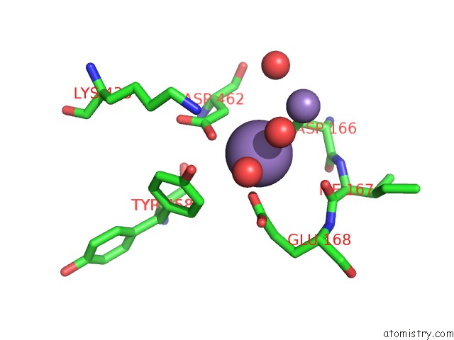
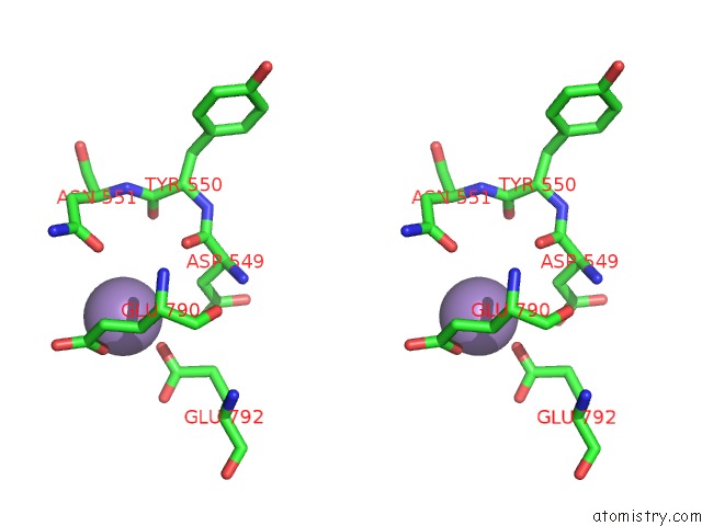
Manganese binding site 1 out of 4 in 5n2g
Go back to
Manganese binding site 1 out
of 4 in the Structure of the E9 Dna Polymerase From Vaccinia Virus in Complex with Manganese
Mono view
Stereo pair view
Mono view
Stereo pair view
A full contact list of Manganese with other atoms in the Mn binding
site number 1 of Structure of the E9 Dna Polymerase From Vaccinia Virus in Complex with Manganese within 5.0Å range:
|
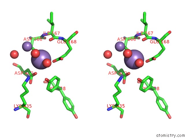
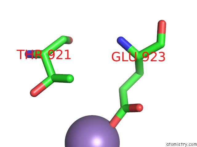
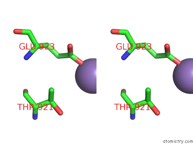
Manganese binding site 2 out of 4 in 5n2g
Go back to
Manganese binding site 2 out
of 4 in the Structure of the E9 Dna Polymerase From Vaccinia Virus in Complex with Manganese
Mono view
Stereo pair view
Mono view
Stereo pair view
A full contact list of Manganese with other atoms in the Mn binding
site number 2 of Structure of the E9 Dna Polymerase From Vaccinia Virus in Complex with Manganese within 5.0Å range:
|
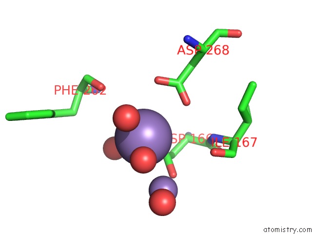
Manganese binding site 3 out of 4 in 5n2g
Go back to
Manganese binding site 3 out
of 4 in the Structure of the E9 Dna Polymerase From Vaccinia Virus in Complex with Manganese

Mono view
Stereo pair view
Mono view
Stereo pair view
A full contact list of Manganese with other atoms in the Mn binding
site number 3 of Structure of the E9 Dna Polymerase From Vaccinia Virus in Complex with Manganese within 5.0Å range:
|
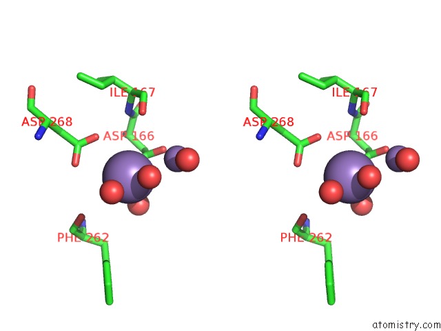
Manganese binding site 4 out of 4 in 5n2g
Go back to
Manganese binding site 4 out
of 4 in the Structure of the E9 Dna Polymerase From Vaccinia Virus in Complex with Manganese
Mono view
Stereo pair view
Mono view
Stereo pair view
A full contact list of Manganese with other atoms in the Mn binding
site number 4 of Structure of the E9 Dna Polymerase From Vaccinia Virus in Complex with Manganese within 5.0Å range:
|
Reference:
N.Tarbouriech,
C.Ducournau,
S.Hutin,
P.J.Mas,
P.Man,
E.Forest,
D.J.Hart,
C.N.Peyrefitte,
W.P.Burmeister,
F.Iseni.
The Vaccinia Virus Dna Polymerase Structure Provides Insights Into the Mode of Processivity Factor Binding. Nat Commun V. 8 1455 2017.
ISSN: ESSN 2041-1723
PubMed: 29129932
DOI: 10.1038/S41467-017-01542-Z
Page generated: Sat Aug 16 18:36:02 2025
ISSN: ESSN 2041-1723
PubMed: 29129932
DOI: 10.1038/S41467-017-01542-Z
Last articles
Na in 6UBHNa in 6UBE
Na in 6UAI
Na in 6UAO
Na in 6UAQ
Na in 6U8T
Na in 6U7P
Na in 6U9J
Na in 6U98
Na in 6U7O