Manganese »
PDB 5hzz-5jpf »
5iwf »
Manganese in PDB 5iwf: Linked KDM5A Jmj Domain Bound to the Inhibitor 2-(((2-((2- (Dimethylamino)Ethyl)(Ethyl)Amino)-2-Oxoethyl)Amino)Methyl) Isonicotinamid
Protein crystallography data
The structure of Linked KDM5A Jmj Domain Bound to the Inhibitor 2-(((2-((2- (Dimethylamino)Ethyl)(Ethyl)Amino)-2-Oxoethyl)Amino)Methyl) Isonicotinamid, PDB code: 5iwf
was solved by
J.R.Horton,
X.Cheng,
with X-Ray Crystallography technique. A brief refinement statistics is given in the table below:
| Resolution Low / High (Å) | 32.99 / 2.29 |
| Space group | C 1 2 1 |
| Cell size a, b, c (Å), α, β, γ (°) | 117.067, 61.918, 46.751, 90.00, 92.28, 90.00 |
| R / Rfree (%) | 19.2 / 24.4 |
Manganese Binding Sites:
The binding sites of Manganese atom in the Linked KDM5A Jmj Domain Bound to the Inhibitor 2-(((2-((2- (Dimethylamino)Ethyl)(Ethyl)Amino)-2-Oxoethyl)Amino)Methyl) Isonicotinamid
(pdb code 5iwf). This binding sites where shown within
5.0 Angstroms radius around Manganese atom.
In total only one binding site of Manganese was determined in the Linked KDM5A Jmj Domain Bound to the Inhibitor 2-(((2-((2- (Dimethylamino)Ethyl)(Ethyl)Amino)-2-Oxoethyl)Amino)Methyl) Isonicotinamid, PDB code: 5iwf:
In total only one binding site of Manganese was determined in the Linked KDM5A Jmj Domain Bound to the Inhibitor 2-(((2-((2- (Dimethylamino)Ethyl)(Ethyl)Amino)-2-Oxoethyl)Amino)Methyl) Isonicotinamid, PDB code: 5iwf:
Manganese binding site 1 out of 1 in 5iwf
Go back to
Manganese binding site 1 out
of 1 in the Linked KDM5A Jmj Domain Bound to the Inhibitor 2-(((2-((2- (Dimethylamino)Ethyl)(Ethyl)Amino)-2-Oxoethyl)Amino)Methyl) Isonicotinamid

Mono view
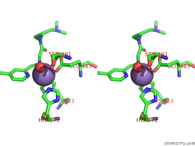
Stereo pair view
Mono view
Stereo pair view
A full contact list of Manganese with other atoms in the Mn binding
site number 1 of Linked KDM5A Jmj Domain Bound to the Inhibitor 2-(((2-((2- (Dimethylamino)Ethyl)(Ethyl)Amino)-2-Oxoethyl)Amino)Methyl) Isonicotinamid within 5.0Å range:
|
Reference:
J.R.Horton,
X.Liu,
M.Gale,
L.Wu,
J.R.Shanks,
X.Zhang,
P.J.Webber,
J.S.Bell,
S.C.Kales,
B.T.Mott,
G.Rai,
D.J.Jansen,
M.J.Henderson,
D.J.Urban,
M.D.Hall,
A.Simeonov,
D.J.Maloney,
M.A.Johns,
H.Fu,
A.Jadhav,
P.M.Vertino,
Q.Yan,
X.Cheng.
Structural Basis For KDM5A Histone Lysine Demethylase Inhibition By Diverse Compounds. Cell Chem Biol V. 23 769 2016.
ISSN: ESSN 2451-9456
PubMed: 27427228
DOI: 10.1016/J.CHEMBIOL.2016.06.006
Page generated: Sat Aug 16 18:13:55 2025
ISSN: ESSN 2451-9456
PubMed: 27427228
DOI: 10.1016/J.CHEMBIOL.2016.06.006
Last articles
Ni in 5FPZNi in 5FPX
Ni in 5FM8
Ni in 5FLH
Ni in 5FLE
Ni in 5FJH
Ni in 5FJK
Ni in 5F5I
Ni in 5ERL
Ni in 5FG6