Atomistry » Manganese » PDB 5gjc-5hwu » 5hrm
Atomistry »
  Manganese »
    PDB 5gjc-5hwu »
      5hrm »

Manganese in PDB 5hrm: Crystal Structure of Phosphotriesterase From Sphingobium Sp. TCM1

Protein crystallography data

The structure of Crystal Structure of Phosphotriesterase From Sphingobium Sp. TCM1, PDB code: 5hrm was solved by M.F.Mabanglo, F.M.Raushel, with X-Ray Crystallography technique. A brief refinement statistics is given in the table below:

Resolution Low / High (Å) 28.93 / 2.05
Space group P 1
Cell size a, b, c (Å), α, β, γ (°) 60.977, 91.273, 96.328, 78.90, 73.60, 89.86
R / Rfree (%) 17.4 / 20.9

Manganese Binding Sites:

The binding sites of Manganese atom in the Crystal Structure of Phosphotriesterase From Sphingobium Sp. TCM1 (pdb code 5hrm). This binding sites where shown within 5.0 Angstroms radius around Manganese atom.
In total 8 binding sites of Manganese where determined in the Crystal Structure of Phosphotriesterase From Sphingobium Sp. TCM1, PDB code: 5hrm:
Jump to Manganese binding site number: 1; 2; 3; 4; 5; 6; 7; 8;

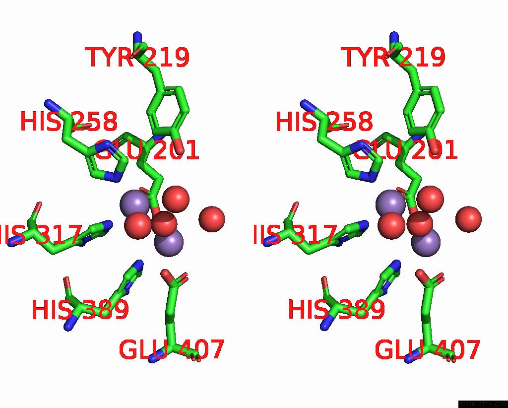
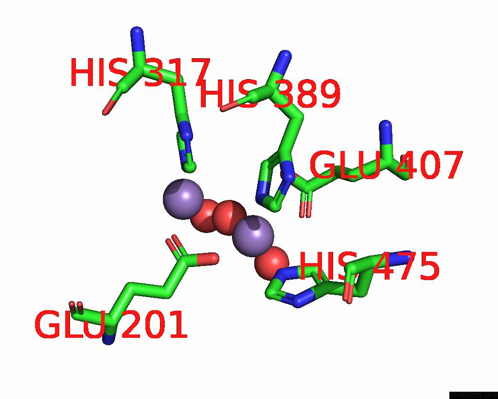
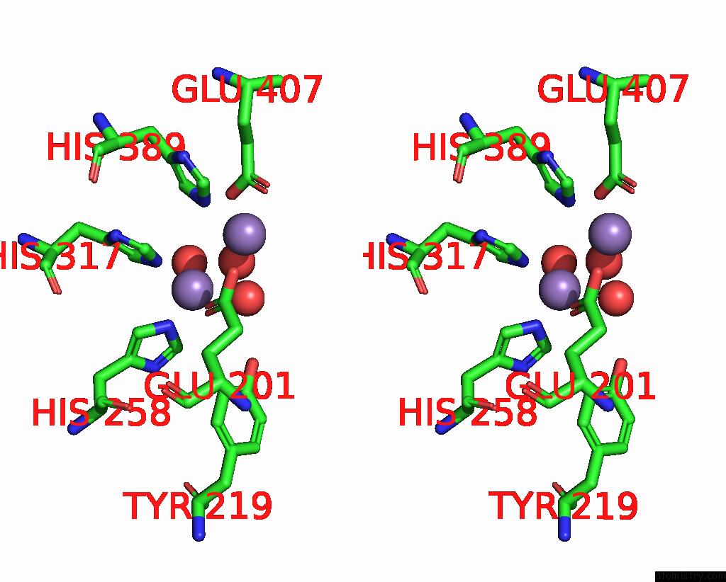
Manganese binding site 1 out of 8 in 5hrm

Go back to Manganese Binding Sites List in 5hrm
Manganese binding site 1 out of 8 in the Crystal Structure of Phosphotriesterase From Sphingobium Sp. TCM1


Mono view


Stereo pair view

A full contact list of Manganese with other atoms in the Mn binding site number 1 of Crystal Structure of Phosphotriesterase From Sphingobium Sp. TCM1 within 5.0Å range:
probe atom residue distance (Å) B Occ
A:Mn601

b:33.1
occ:1.00
OE2 A:GLU201 2.1 33.7 1.0
O A:HOH864 2.1 38.4 1.0
NE2 A:HIS475 2.1 28.5 1.0
NE2 A:HIS389 2.3 29.5 1.0
OE1 A:GLU407 2.6 40.5 1.0
O A:HOH932 3.0 49.6 1.0
CE1 A:HIS475 3.0 32.7 1.0
CD2 A:HIS475 3.0 30.8 1.0
CD A:GLU201 3.1 33.6 1.0
CE1 A:HIS389 3.1 30.9 1.0
CD2 A:HIS389 3.3 28.6 1.0
CD A:GLU407 3.4 40.6 1.0
OE1 A:GLU201 3.5 31.6 1.0
MN A:MN602 3.5 31.0 1.0
OE2 A:GLU407 3.6 38.2 1.0
NE2 A:HIS317 4.0 32.9 1.0
ND1 A:HIS475 4.1 28.3 1.0
CE1 A:HIS317 4.1 30.9 1.0
CG A:HIS475 4.1 32.4 1.0
ND1 A:HIS389 4.3 31.9 1.0
CG A:GLU201 4.3 30.8 1.0
CG A:HIS389 4.3 28.1 1.0
O A:HOH858 4.5 49.1 1.0
CG A:GLU407 4.6 34.4 1.0
CB A:GLU407 4.7 30.1 1.0

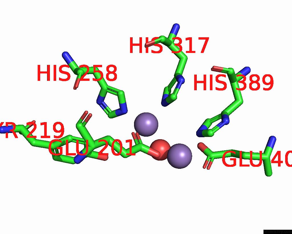
Manganese binding site 2 out of 8 in 5hrm

Go back to Manganese Binding Sites List in 5hrm
Manganese binding site 2 out of 8 in the Crystal Structure of Phosphotriesterase From Sphingobium Sp. TCM1


Mono view


Stereo pair view

A full contact list of Manganese with other atoms in the Mn binding site number 2 of Crystal Structure of Phosphotriesterase From Sphingobium Sp. TCM1 within 5.0Å range:
probe atom residue distance (Å) B Occ
A:Mn602

b:31.0
occ:1.00
OE1 A:GLU201 2.0 31.6 1.0
NE2 A:HIS317 2.0 32.9 1.0
O A:HOH864 2.1 38.4 1.0
NE2 A:HIS258 2.1 31.9 1.0
CD2 A:HIS317 2.9 27.8 1.0
CD A:GLU201 3.0 33.6 1.0
CD2 A:HIS258 3.0 34.3 1.0
O A:HOH858 3.0 49.1 1.0
CE1 A:HIS317 3.1 30.9 1.0
CE1 A:HIS258 3.1 30.9 1.0
O A:HOH939 3.4 50.6 1.0
OE2 A:GLU201 3.4 33.7 1.0
MN A:MN601 3.5 33.1 1.0
CG A:HIS317 4.1 28.3 1.0
ND1 A:HIS317 4.1 31.5 1.0
CG A:HIS258 4.2 34.0 1.0
ND1 A:HIS258 4.2 29.6 1.0
OH A:TYR219 4.2 36.3 1.0
NE2 A:HIS389 4.3 29.5 1.0
CG A:GLU201 4.3 30.8 1.0
CE2 A:TYR219 4.5 34.7 1.0
CD2 A:HIS389 4.5 28.6 1.0
OE2 A:GLU407 4.5 38.2 1.0
O A:HOH932 4.8 49.6 1.0
CB A:GLU201 4.8 31.6 1.0
CZ A:TYR219 4.9 34.8 1.0

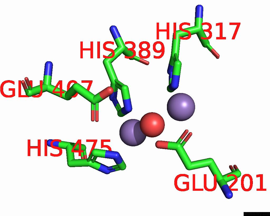
Manganese binding site 3 out of 8 in 5hrm

Go back to Manganese Binding Sites List in 5hrm
Manganese binding site 3 out of 8 in the Crystal Structure of Phosphotriesterase From Sphingobium Sp. TCM1


Mono view


Stereo pair view

A full contact list of Manganese with other atoms in the Mn binding site number 3 of Crystal Structure of Phosphotriesterase From Sphingobium Sp. TCM1 within 5.0Å range:
probe atom residue distance (Å) B Occ
B:Mn601

b:28.1
occ:1.00
OE1 B:GLU201 2.0 20.0 1.0
NE2 B:HIS258 2.1 27.6 1.0
O B:HOH712 2.1 38.4 1.0
NE2 B:HIS317 2.2 28.4 1.0
CD2 B:HIS258 2.9 29.8 1.0
CD2 B:HIS317 3.0 23.2 1.0
CD B:GLU201 3.0 20.0 1.0
CE1 B:HIS258 3.2 29.4 1.0
CE1 B:HIS317 3.3 31.0 1.0
OE2 B:GLU201 3.3 20.0 1.0
MN B:MN602 3.6 31.1 1.0
CG B:HIS258 4.1 29.4 1.0
CG B:HIS317 4.2 29.3 1.0
OH B:TYR219 4.2 35.1 1.0
ND1 B:HIS258 4.2 30.2 1.0
ND1 B:HIS317 4.3 27.0 1.0
NE2 B:HIS389 4.3 30.9 1.0
CG B:GLU201 4.3 20.0 1.0
CE2 B:TYR219 4.4 28.2 1.0
CD2 B:HIS389 4.6 28.0 1.0
OE2 B:GLU407 4.6 39.2 1.0
CB B:GLU201 4.7 20.0 1.0
CZ B:TYR219 4.8 34.3 1.0

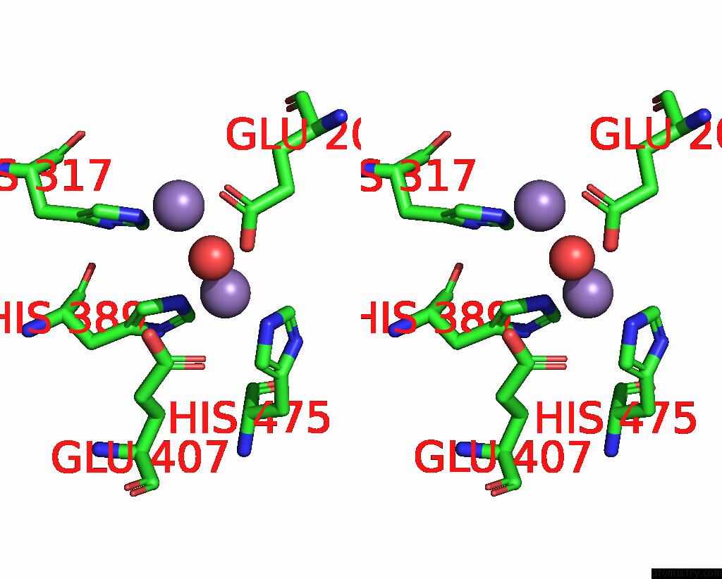
Manganese binding site 4 out of 8 in 5hrm

Go back to Manganese Binding Sites List in 5hrm
Manganese binding site 4 out of 8 in the Crystal Structure of Phosphotriesterase From Sphingobium Sp. TCM1


Mono view


Stereo pair view

A full contact list of Manganese with other atoms in the Mn binding site number 4 of Crystal Structure of Phosphotriesterase From Sphingobium Sp. TCM1 within 5.0Å range:
probe atom residue distance (Å) B Occ
B:Mn602

b:31.1
occ:1.00
OE2 B:GLU201 1.8 20.0 1.0
O B:HOH712 2.1 38.4 1.0
NE2 B:HIS475 2.1 25.2 1.0
NE2 B:HIS389 2.2 30.9 1.0
OE1 B:GLU407 2.7 38.0 1.0
CD B:GLU201 2.9 20.0 1.0
CD2 B:HIS475 3.0 25.6 1.0
CE1 B:HIS389 3.1 29.8 1.0
CE1 B:HIS475 3.1 27.9 1.0
CD2 B:HIS389 3.2 28.0 1.0
CD B:GLU407 3.4 35.5 1.0
OE1 B:GLU201 3.5 20.0 1.0
OE2 B:GLU407 3.6 39.2 1.0
MN B:MN601 3.6 28.1 1.0
NE2 B:HIS317 4.0 28.4 1.0
CG B:HIS475 4.2 24.9 1.0
CE1 B:HIS317 4.2 31.0 1.0
ND1 B:HIS475 4.2 27.6 1.0
ND1 B:HIS389 4.2 26.9 1.0
CG B:GLU201 4.2 20.0 1.0
CG B:HIS389 4.3 27.8 1.0
CG B:GLU407 4.7 29.6 1.0
CB B:GLU407 4.8 29.9 1.0
CZ B:PHE476 5.0 33.7 1.0

Manganese binding site 5 out of 8 in 5hrm

Go back to Manganese Binding Sites List in 5hrm
Manganese binding site 5 out of 8 in the Crystal Structure of Phosphotriesterase From Sphingobium Sp. TCM1


Mono view


Stereo pair view

A full contact list of Manganese with other atoms in the Mn binding site number 5 of Crystal Structure of Phosphotriesterase From Sphingobium Sp. TCM1 within 5.0Å range:
probe atom residue distance (Å) B Occ
C:Mn601

b:30.1
occ:1.00
OE1 C:GLU201 2.0 36.1 1.0
NE2 C:HIS317 2.1 30.8 1.0
NE2 C:HIS258 2.1 31.5 1.0
O C:HOH803 2.2 42.4 1.0
CD C:GLU201 3.0 36.5 1.0
CD2 C:HIS317 3.0 27.8 1.0
CD2 C:HIS258 3.1 31.7 1.0
CE1 C:HIS317 3.1 29.0 1.0
CE1 C:HIS258 3.2 31.2 1.0
OE2 C:GLU201 3.3 31.7 1.0
MN C:MN602 3.6 31.0 1.0
ND1 C:HIS317 4.2 30.7 1.0
CG C:HIS317 4.2 29.8 1.0
OH C:TYR219 4.2 38.4 1.0
ND1 C:HIS258 4.2 34.2 1.0
CG C:HIS258 4.2 33.9 1.0
NE2 C:HIS389 4.3 33.8 1.0
CG C:GLU201 4.3 35.4 1.0
CE2 C:TYR219 4.5 32.8 1.0
CD2 C:HIS389 4.5 31.0 1.0
OE2 C:GLU407 4.6 39.4 1.0
CB C:GLU201 4.8 34.5 1.0
CZ C:TYR219 4.8 35.9 1.0

Manganese binding site 6 out of 8 in 5hrm

Go back to Manganese Binding Sites List in 5hrm
Manganese binding site 6 out of 8 in the Crystal Structure of Phosphotriesterase From Sphingobium Sp. TCM1


Mono view


Stereo pair view

A full contact list of Manganese with other atoms in the Mn binding site number 6 of Crystal Structure of Phosphotriesterase From Sphingobium Sp. TCM1 within 5.0Å range:
probe atom residue distance (Å) B Occ
C:Mn602

b:31.0
occ:1.00
OE2 C:GLU201 2.0 31.7 1.0
NE2 C:HIS475 2.1 26.9 1.0
O C:HOH803 2.1 42.4 1.0
NE2 C:HIS389 2.2 33.8 1.0
OE1 C:GLU407 2.7 41.5 1.0
CD2 C:HIS475 3.0 31.1 1.0
CD C:GLU201 3.1 36.5 1.0
CE1 C:HIS475 3.1 30.6 1.0
CE1 C:HIS389 3.1 31.9 1.0
CD2 C:HIS389 3.2 31.0 1.0
CD C:GLU407 3.4 41.7 1.0
OE2 C:GLU407 3.5 39.4 1.0
OE1 C:GLU201 3.5 36.1 1.0
MN C:MN601 3.6 30.1 1.0
NE2 C:HIS317 4.1 30.8 1.0
ND1 C:HIS475 4.1 30.0 1.0
CG C:HIS475 4.1 32.9 1.0
CE1 C:HIS317 4.1 29.0 1.0
ND1 C:HIS389 4.2 31.8 1.0
CG C:HIS389 4.3 31.6 1.0
CG C:GLU201 4.3 35.4 1.0
CG C:GLU407 4.7 35.6 1.0
CB C:GLU407 4.9 33.4 1.0

Manganese binding site 7 out of 8 in 5hrm

Go back to Manganese Binding Sites List in 5hrm
Manganese binding site 7 out of 8 in the Crystal Structure of Phosphotriesterase From Sphingobium Sp. TCM1


Mono view


Stereo pair view

A full contact list of Manganese with other atoms in the Mn binding site number 7 of Crystal Structure of Phosphotriesterase From Sphingobium Sp. TCM1 within 5.0Å range:
probe atom residue distance (Å) B Occ
D:Mn601

b:35.6
occ:1.00
OE2 D:GLU201 2.0 31.0 1.0
NE2 D:HIS475 2.1 31.3 1.0
NE2 D:HIS389 2.2 31.1 1.0
O D:HOH858 2.2 43.3 1.0
OE1 D:GLU407 2.6 43.1 1.0
CE1 D:HIS475 3.0 36.1 1.0
CD2 D:HIS475 3.0 30.9 1.0
CD D:GLU201 3.0 33.6 1.0
CE1 D:HIS389 3.1 32.1 1.0
O D:HOH864 3.2 57.5 1.0
CD2 D:HIS389 3.2 28.6 1.0
CD D:GLU407 3.4 42.8 1.0
OE1 D:GLU201 3.4 32.2 1.0
MN D:MN602 3.6 35.0 1.0
OE2 D:GLU407 3.6 42.0 1.0
NE2 D:HIS317 4.0 34.4 1.0
ND1 D:HIS475 4.1 32.1 1.0
CG D:HIS475 4.1 34.5 1.0
CE1 D:HIS317 4.2 33.2 1.0
ND1 D:HIS389 4.2 33.6 1.0
CG D:HIS389 4.3 31.1 1.0
CG D:GLU201 4.3 37.9 1.0
CG D:GLU407 4.7 35.6 1.0
CB D:GLU407 4.9 31.4 1.0
O D:HOH838 4.9 52.8 1.0

Manganese binding site 8 out of 8 in 5hrm

Go back to Manganese Binding Sites List in 5hrm
Manganese binding site 8 out of 8 in the Crystal Structure of Phosphotriesterase From Sphingobium Sp. TCM1


Mono view


Stereo pair view

A full contact list of Manganese with other atoms in the Mn binding site number 8 of Crystal Structure of Phosphotriesterase From Sphingobium Sp. TCM1 within 5.0Å range:
probe atom residue distance (Å) B Occ
D:Mn602

b:35.0
occ:1.00
OE1 D:GLU201 2.0 32.2 1.0
NE2 D:HIS258 2.0 33.9 1.0
NE2 D:HIS317 2.0 34.4 1.0
O D:HOH858 2.2 43.3 1.0
CD2 D:HIS317 2.9 31.0 1.0
CD2 D:HIS258 2.9 35.0 1.0
O D:HOH878 3.0 53.4 1.0
CE1 D:HIS258 3.1 31.4 1.0
CD D:GLU201 3.1 33.6 1.0
CE1 D:HIS317 3.1 33.2 1.0
O D:HOH838 3.3 52.8 1.0
OE2 D:GLU201 3.4 31.0 1.0
MN D:MN601 3.6 35.6 1.0
CG D:HIS317 4.1 36.1 1.0
CG D:HIS258 4.1 35.1 1.0
ND1 D:HIS258 4.1 33.0 1.0
ND1 D:HIS317 4.1 31.5 1.0
NE2 D:HIS389 4.3 31.1 1.0
OH D:TYR219 4.4 39.7 1.0
CG D:GLU201 4.4 37.9 1.0
CD2 D:HIS389 4.5 28.6 1.0
CE1 D:TYR219 4.5 35.4 1.0
OE2 D:GLU407 4.6 42.0 1.0
CB D:GLU201 4.9 32.9 1.0
CZ D:TYR219 5.0 37.9 1.0

Reference:

M.F.Mabanglo, D.F.Xiang, A.N.Bigley, F.M.Raushel. Structure of A Novel Phosphotriesterase From Sphingobium Sp. TCM1: A Familiar Binuclear Metal Center Embedded in A Seven-Bladed Beta-Propeller Protein Fold. Biochemistry V. 55 3963 2016.
ISSN: ISSN 0006-2960
PubMed: 27353520
DOI: 10.1021/ACS.BIOCHEM.6B00364
Page generated: Sat Aug 16 17:39:46 2025

Last articles

Mn in 9LJU
Mn in 9LJW
Mn in 9LJS
Mn in 9LJR
Mn in 9LJT
Mn in 9LJV
Mg in 9UA2
Mg in 9R96
Mg in 9VM1
Mg in 9P01
© Copyright 2008-2020 by atomistry.com
Home   |    Site Map   |    Copyright   |    Contact us   |    Privacy