Manganese »
PDB 5ekw-5fxv »
5f56 »
Manganese in PDB 5f56: Structure of Recj Complexed with Dna and Ssb-Ct
Protein crystallography data
The structure of Structure of Recj Complexed with Dna and Ssb-Ct, PDB code: 5f56
was solved by
Y.Zhao,
Y.Hua,
K.Cheng,
with X-Ray Crystallography technique. A brief refinement statistics is given in the table below:
| Resolution Low / High (Å) | 29.05 / 2.30 |
| Space group | P 32 2 1 |
| Cell size a, b, c (Å), α, β, γ (°) | 102.220, 102.220, 166.120, 90.00, 90.00, 120.00 |
| R / Rfree (%) | 22.6 / 23.9 |
Manganese Binding Sites:
The binding sites of Manganese atom in the Structure of Recj Complexed with Dna and Ssb-Ct
(pdb code 5f56). This binding sites where shown within
5.0 Angstroms radius around Manganese atom.
In total 2 binding sites of Manganese where determined in the Structure of Recj Complexed with Dna and Ssb-Ct, PDB code: 5f56:
Jump to Manganese binding site number: 1; 2;
In total 2 binding sites of Manganese where determined in the Structure of Recj Complexed with Dna and Ssb-Ct, PDB code: 5f56:
Jump to Manganese binding site number: 1; 2;
Manganese binding site 1 out of 2 in 5f56
Go back to
Manganese binding site 1 out
of 2 in the Structure of Recj Complexed with Dna and Ssb-Ct
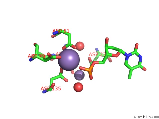
Mono view
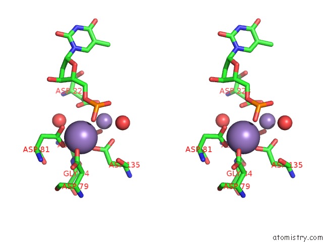
Stereo pair view
Mono view
Stereo pair view
A full contact list of Manganese with other atoms in the Mn binding
site number 1 of Structure of Recj Complexed with Dna and Ssb-Ct within 5.0Å range:
|
Manganese binding site 2 out of 2 in 5f56
Go back to
Manganese binding site 2 out
of 2 in the Structure of Recj Complexed with Dna and Ssb-Ct
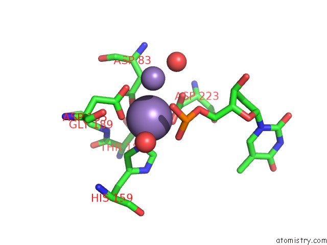
Mono view
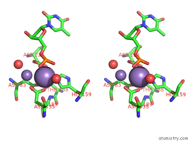
Stereo pair view
Mono view
Stereo pair view
A full contact list of Manganese with other atoms in the Mn binding
site number 2 of Structure of Recj Complexed with Dna and Ssb-Ct within 5.0Å range:
|
Reference:
K.Cheng,
H.Xu,
X.Chen,
L.Wang,
B.Tian,
Y.Zhao,
Y.Hua.
Structural Basis For Dna 5 -End Resection By Recj Elife V. 5 14294 2016.
ISSN: ESSN 2050-084X
PubMed: 27058167
DOI: 10.7554/ELIFE.14294
Page generated: Sun Oct 6 00:11:20 2024
ISSN: ESSN 2050-084X
PubMed: 27058167
DOI: 10.7554/ELIFE.14294
Last articles
K in 3I6TK in 3I56
K in 3I55
K in 3HQO
K in 3I4D
K in 3HW9
K in 3HRZ
K in 3HPL
K in 3H8G
K in 3HQN